Radiologists Can Deftly Curb Motion Artifacts in Abdominal MRI
Motion mitigation is critically important to improve image quality and workflow efficiency

Diagnostic-quality T1-weighted (T1W) images are an important component of standard MRI scans for the abdomen. Most MRI procedures include a single-phase or multi-phase series of these images, but they can be affected by patient motion---especially when the patients have trouble with breath-holding---and can compromise the image quality and exam efficiency.
In a RadioGraphics article, radiologists and physicists from academia and industry discuss methods to reduce motion artefact during scans by using specialized techniques for capturing images. “Some of the challenges in abdominal MRI are to predict how well patients will be able to hold their breath during the exam and how to select the best motion mitigation strategy,” said lead author Orpheus Kolokythas, MD, professor of radiology and director of MRI at the University of Washington in Seattle.
Radiologists should consider factors like the patient’s condition, the clinical problem they’re investigating, and the technical ramifications of the method for an effective acquisition. In their RadioGraphics article, Dr. Kolokythas and colleagues explore the technical principles, commercial availability, clinical applications and limitations of various ways to mitigate motion.

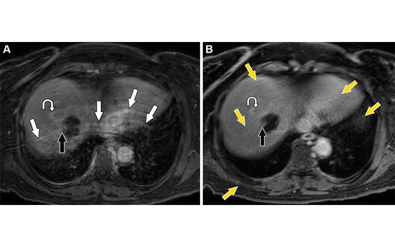
Comparison of postcontrast Cartesian BH and radial k-space free-breathing techniques during the equilibrium phase in a 63-year-old woman 6 months after radiofrequency ablation of hepatocellular carcinoma in the right hepatic lobe. (A) Axial contrast-enhanced Cartesian 3D T1W Dixon image with poor patient BH shows curvilinear aliasing artifacts (white arrows) that raise the question of nodular enhancement at the margin of the treatment zone, suggestive of residual tumor (black arrow). (B) Axial free-breathing radial k-space image, which required 3 minutes and 10 seconds of additional imaging, was performed immediately after the BH series. The curved arrow in A and B denotes a peripherally enhancing satellite nodule with washout (site of hepatocellular carcinoma recurrence), which is better seen on the radial image. Motion artifacts in the form of streaks are present in B (yellow arrows). However, these artifacts are less impactful than the aliasing artifacts seen in A. The suspicious finding at the margin of the treatment zone on the Cartesian image represents a small portion of nonneoplastic hepatic tissue (black arrow). Also, note the overall increased blur in B compared with that in A. 3-T BH GRE Dixon imaging parameters: TR, 3.5 msec; TE, 1.32/2.3 msec; flip angle, 10°; section thickness, 3.2 mm; acquisition matrix, 200 × 200. 3-T free-breathing GRE Dixon radial imaging parameters: TR, 4.8 msec; TE, 1.4/2.7 msec; flip angle, 10°; section thickness, 3.2 mm; acquisition matrix, 300 × 572. https://doi.org/10.1148/rg.230173 © RSNA 2024
Respiratory Gating and Triggering
Non-phasic motions, such as hollow viscus peristalsis or patient movement, cannot be predicted. But to work with the comparatively predictable motions of breathing and arterial pulsation, gating and triggering techniques selectively capture data while letting the patient breathe freely.
With respiratory gating, data are captured as a change in MRI signal and translated into a signal acquisition when the patient is exhaling. Information outside the expiration phase is discarded.
Respiratory triggering employs external sensors, such as cameras and bellows placed on the lower chest or upper abdomen to pick up breathing motion.
“Since both gated and triggered techniques rely on consistent breathing, coaching the patient before and during the exam is of utmost importance,” Dr. Kolokythas said. “Technologists should explain what to expect during the exam, give simple and clear instructions for how to breathe in the upcoming sequence and explain to the patient that regular consistent breathing will help shorten exam time and optimize images.”
Nondynamic Radial k-Space Sampling
Conventional Cartesian k-space readouts are subject to discrete ghost artifacts after reconstruction, because motion during the examination registers at a different location along the y-axis. To offset these artifacts, radiologists can select trajectories that sample the phase-encoding direction in a radial spoke pattern.
“Golden angle radial sampling is a specific radial profile that uses nonuniform distribution of angles between radial lines,” the authors explained. “In contrast to sequential ordering of radial spokes, golden angle sampling helps ensure homogenous filling of k-space at any given point throughout the acquisition cycle.” This bolsters the motion mitigation effect of T1 radial sampling schemes.
The “stack-of-stars” technique employs a stack of two-dimensional sampling profiles in the z-direction to acquire volumetric k-space data in all three spatial dimensions. Combining golden angle and stack-of-stars sequences yields 3D data with higher motion robustness and more protocol flexibility than Cartesian k-space sampling methods.
Moving Forward
Exciting future applications are on the horizon as well, the authors noted. For example, a deep convolutional network, which is a type of artificial neural network specifically designed for image analysis, could be trained to extract artificially generated noise over images generated with inadequate breath holds, thereby successfully decreasing motion and blurring that resulted from a free-breathing Cartesian sampling. Meanwhile, 3D trajectories under investigation in the cardiac imaging realm could be useful in MRI applications that require isotropic high resolution, or a 3D image where the spatial resolution is the same in all directions.
Dr. Kolokythas reiterated that it is of paramount importance to coach the patient in the MRI suite.
“Motion correction and mitigation techniques can help improve image quality and should be implemented, where available, in a way so they can be seamlessly applied,” he said. “To save overall examination time, a brief clinical breath-hold assessment can be performed on alert patients outside the MRI suite to confirm inability for adequate breath holds. This assessment provides valuable information to the MRI technologist, who can use it to choose the appropriate examination strategy from the beginning.”
For More Information
Access the RadioGraphics article, “T1-weighted Motion Mitigation in Abdominal MRI: Technical Principles, Clinical Applications, Current Limitations, and Future Prospects.”
Read previous RSNA News stories about the use of abdominal MRI: