Somatostatin Receptor PET/MRI Useful For Imaging Head And Neck Cancers
Improved soft tissue evaluation assists in imaging the small spaces and complex anatomy of the head and neck

In concert with the RadioGraphics monograph, RSNA News shares the latest in head and neck imaging.
Hybrid imaging with PET/MRI and novel radiotracers is poised to revolutionize the diagnosis and treatment of neuroendocrine tumors of the head and neck, offering superior soft tissue resolution and improvements in radiation therapy among other benefits.
Somatostatin receptor-expressing tumors can develop in many parts of the body. In the head and neck, they include paragangliomas and meningiomas, as well as some pituitary tumors, olfactory neuroblastomas, middle ear neuroendocrine tumors, and medullary thyroid cancer. Many of these lesions are difficult to detect and diagnose with routine structural imaging.
Development of somatostatin receptor (SSTR) radiopharmaceuticals, including the commonly used gallium-68 and copper-64 DOTATATE, has opened promising options for diagnosis and treatment of somatostatin receptor-expressing tumors. DOTATATE derives its name from a short-chain peptide analogous to the hormone somatostatin. Somatostatin receptors on the surface of the cell take up the tracer. Many neuroendocrine tumors take up somatostatin more avidly than normal cells, and this uptake is visible on PET imaging.
PET is typically paired with CT for better anatomical information, but in recent years the nuclear imaging modality has been used with MRI for improved soft tissue visualization. The potential of this hybrid imaging is vast, according to researchers who recently published a RadioGraphics review of its benefits.
“We want to spread awareness of hybrid PET/MRI because we think it should be more widely utilized,” said lead author Graham Keir, MD, neuroradiology fellow at Weill Cornell, New York Presbyterian Hospital in New York City. “From our experience, it’s a powerful imaging modality that has numerous applications.”
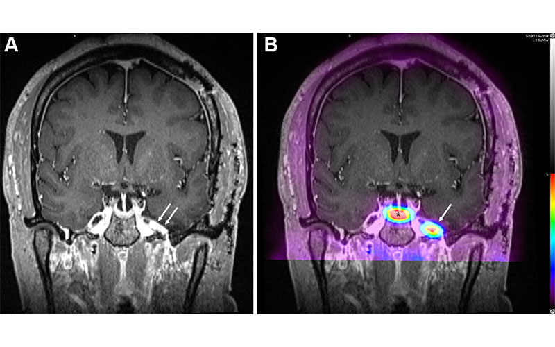
Residual or recurrent meningioma in a 38-year-old woman who presented with concern for recurrence after undergoing left pterional craniotomy and resection of a meningioma. (A) Postoperative coronal contrast-enhanced T1-weighted MR image shows prominent enhancement (arrows) along the anteromedial left temporal convexity measuring up to 1.3 cm, approximating the inferolateral aspect of the Meckel cave and bordering the left carotid artery. (B) Fused coronal image from DOTATATE PET/MRI shows DOTATATE avidity in this region (arrow) with a maximum standardized uptake value (SUVmax) of 15.6, compatible with residual or recurrent meningioma. Note the physiologic uptake of DOTATATE by the normal pituitary tissue (*). https://doi.org/10.1148/rg.240020 © RSNA 2024
Improving Lesion Localization, Disease Surveillance and Radiation Therapy Planning
PET/MRI with DOTATATE has several key advantages over somatostatin receptor scintigraphy for imaging the small spaces and complex anatomy of the head and neck. It has higher sensitivity for lesion detection while offering lower radiation doses and shorter exam times.
For meningiomas, it is excellent at evaluating the extent of disease and distinguishing it from similar-appearing lesions like schwannoma.
“This is helpful because for example in the orbit if you see a lesion and you’re having trouble distinguishing between a meningioma versus a non-somatostatin receptor-expressing orbital lesion, if you see that tracer uptake you can be more confident that it’s the former. Similarly, SSTR-PET/MRI allows us to distinguish cerebellopontine angle meningiomas from schwannomas,” Dr. Keir said.
Some studies have shown that the more precise localization of lesions in the treatment of meningiomas results in a lower radiation dose to critical structures like the optic nerve. Somatostatin receptor PET/MRI enables more accurate radiation dose contouring and is effective after surgery in distinguishing disease from other post-surgical changes.
“When you have that uptake on PET, it makes you more confident that there is residual or recurrent disease after surgery that needs to be treated,” Dr. Keir said.
DOTATATE’s ability to detect multiple lesions is critical in the setting of head and neck paragangliomas, up to a third of which may be hereditary.
“Multiple neuroendocrine tumors suggest an underlying genetic syndrome,” Dr. Keir said. “That has implications not only for the patient, but also family members who can subsequently get the recommended genetic testing.”
Somatostatin receptor PET/MRI is also helpful for distinguishing paragangliomas from surrounding blood vessels.
In medullary thyroid cancer, the third most common type of thyroid cancer, somatostatin receptor PET/MRI has shown advantages over FDG-PET for sensitivity and incidence of lesion detection. It also has potential roles in the evaluation of middle ear adenomas, pituitary neuroendocrine tumors, and olfactory neuroblastoma.
Somatostatin receptor PET/MRI has limitations. Not all lesions that pick up DOTATATE are neuroendocrine tumors. Many papillary thyroid cancers, for instance, display somatostatin receptor expression. When limited to the head and neck, somatostatin receptor PET/MRI runs the risk of missing metastatic disease elsewhere in the body. Whole-body PET/MRI may be needed if metastatic disease is suspected.
The Intersection of Nuclear Medicine and Neuroradiology
The true potential of somatostatin receptor PET/MRI is limited by the relative scarcity of hybrid PET/MRI scanners. A workaround to this is available in the form of commercial software that allows users to fuse head and neck PET data from a PET/CT scan with recently acquired structural MRI scans.
“There are only a few centers that are using DOTATATE for neuroendocrine head and neck imaging,” Dr. Keir said. “We need more experience and more studies but it’s certainly a very promising modality.”
Dr. Keir expects somatostatin receptor PET/MRI to become a mainstay in the workup of patients as more radiologists and clinicians become aware of its advantages. It has uses in treatment monitoring and decision-making, radiation dose and contouring determinations and decisions on when patients need to go back for surgery.
“The intersection of nuclear medicine and neuroradiology is a really exciting place right now,” Dr. Keir said. “Over time, I think we’ll see somatostatin receptor PET/MRI much more within the clinical workflow as more molecular imaging radiotracers come out that will be specific to different types of cancers.”
For More Information
Access the RadioGraphics paper, “Hybrid Somatostatin Receptor (SSTR) PET/MRI of the Head and Neck.”
Read previous RSNA News stories on head and neck imaging: