Sinonasal Tumors Receive New Classification From WHO
New framework will help radiologists understand the appearance and multidisciplinary considerations for these tumors

In concert with the RadioGraphics monograph, RSNA News shares the latest in head and neck imaging.
Sinonasal tumors are a heterogenous group involving a variety of tissues within the nasal cavity or paranasal sinuses. Although such tumors are relatively rare, representing just three percent of tumors in the upper respiratory tract, they can cause morbidity and mortality for patients.
With nonspecific symptoms such as pain and congestion, patients tend to present with relatively advanced disease. As a result, at diagnosis, the five-year survival rate for localized nasal cavity and paranasal sinus cancer is about 86% and is 52% for regional nasal cavity and paranasal sinus cancer.
“The good news is there’s been an evolution in sinonasal tumor research, the result being an increased awareness of the distinctive molecular and genetic characteristics of tumors in this location,” said Greg Avey, MD, director of head and neck imaging at the University of Wisconsin–Madison.
To this end, in 2022, the World Health Organization (WHO) released a new classification of head and neck malignancies—a classification that emphasizes tumor molecular and genetic characteristics.
“The addition of this new WHO classification scheme provides a unique opportunity to review the imaging appearance of sinonasal tumors, along with important multidisciplinary considerations for common tumor types,” added Dr. Avey, who co-authored a review for RadioGraphics.
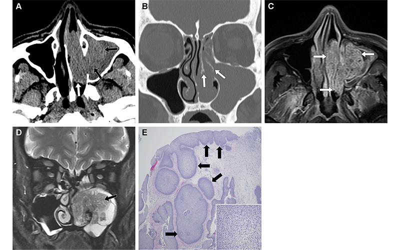
Inverted papilloma in a 66-year-old man who presented with a concern for sinusitis. (A) Axial soft-tissue unenhanced CT image shows a hyperattenuating mass in the left maxillary sinus (black arrows) with extension into the left nasal cavity (white arrow). (B) Coronal unenhanced CT image (bone window) shows hyperostosis of the left middle turbinate (arrows), likely reflecting the osseous attachment of this lesion. (C) Axial contrast-enhanced T1-weighted MR image shows that the mass enhances (arrows) after administration of contrast material, suggesting that this is not an antrochoanal polyp. (D) Coronal T2-weighted MR image shows that the mass has a “cerebriform” appearance (arrow), which is characteristic of an inverted papilloma. This mass was surgically resected. (E) Photomicrograph shows an inverted papillary growth pattern (arrows) with an otherwise flat apical surface. (Hematoxylin-eosin [H-E) stain; objective magnification, ×4.) Inset photomicrograph shows stromal edema with scattered infiltrating neutrophils, which are commonly observed. (H-E stain; objective magnification, ×40.) https://doi.org/10.1148/rg.240035 © RSNA 2024
Imaging’s Role in Managing Sinonasal Tumors
When it comes to managing sinonasal tumors, imaging is primarily used for mapping—a task that involves determining the involvement of the sinonasal cavity subsites, surrounding soft tissues, orbital contents, intracranial compartment, perineural structures and lymph nodes.
“CT and MRI are the preferred initial imaging modalities for evaluating a patient with a new sinonasal mass, with each offering specific advantages and disadvantages,” Dr. Avey explained.
For example, whereas CT excels at detecting bone remodeling and erosion, MRI is better at demonstrating bone marrow involvement. On the other hand, MRI is the modality of choice for differentiating inflammatory secretions from enhancing tumor, as CT may falsely suggest post-obstructed secretions as tumor involvement.
Differentiating malignant tumors from benign entities is another key role of imaging in sinonasal tumors—a process that relies on specific imaging features seen on CT and MRI. For instance, benign conditions typically demonstrate smooth, well-defined margins, displaced tissue planes, bone remodeling, and T2 hyperintense signal on MRI. In contrast, malignant lesions are more likely to have infiltrative margins, invaded tissue planes, bone invasion, and a relatively T2 hypointense signal compared to normal mucosa.
Radiology as Part of a Multidisciplinary Approach
While radiology has a big role to play, both in tumor diagnosis and treatment, this role cannot happen in a vacuum. It must be part of a multidisciplinary approach.
“Our role as radiologists is to recognize these tumors, help delineate their full extent and guide therapy,” Dr. Avey said. “When we recognize complicating elements, such as the tumor’s spread along the cranial nerves, we can help guide the patient to more appropriate therapy and better outcomes.”
The updated WHO classification can help radiologists make such patient-specific assessments.
The new classification separates tumor types that occur more broadly in the body and focuses on those that occur more specifically in the sinonasal region. It also adds several new categories, such as Switch/Sucrose Non-Fermentable (SWI/SNF) deficient carcinomas and DEK-AFF2 carcinomas.
According to Dr. Avey, radiologists must understand these evolving categories to partner more completely with their other colleagues. As an example, he refers to DEK-AFF2 carcinomas, that occur almost exclusively on the posterior nasal turbinate and are more aggressive than expected based on their histopathological appearance.
“By suggesting that a tumor could be a DEK-AFF2 carcinoma, we might prompt a fuller molecular evaluation and additional therapy to reflect the aggressiveness of this tumor,” he said.
Treatment Starts With Recognizing the Tumor
The changes to the WHO classification resulted from a marked shift in health care’s understanding of sinonasal tumors and their prognosis. Eventually, these changes will lead to new, more tailored therapies and, ultimately, better outcomes.
“The tumor board wants us to recognize the tumor first, as many will first be identified via imaging performed for nonspecific symptoms,” Dr. Avey said. “After that, our job is to guide the efficient evaluation of these patients and make sure the treatment team considers the many potential entities that can make up tumors in this region.”
For More Information
Access the RadioGraphics article, “Sinonasal Tumors: What the Multidisciplinary Cancer Care Board Wants to Know.”
Read previous RSNA News stories on head and neck imaging: