Understanding Hearing Loss
The role of imaging in the diagnosis and treatment

In concert with the RadioGraphics monograph, RSNA News shares the latest in head and neck imaging.
Hearing loss affects more than a billion people worldwide. The use of imaging – temporal bone CT scans in particular - alongside results of audiologic tests and knowledge of the physical principles of hearing, has permitted radiologists to be uniquely positioned to contribute to comprehensive and effective treatment plans for patients.
“By incorporating the results of audiologic testing into their evaluation, radiologists can perform a more informed and more intentional search for the structural cause of hearing loss,” said William Malouf, MD, associate professor of radiology at Wake Forest University, Winston-Salem, NC, and an author of a RadioGraphics article about the multidisciplinary approach to diagnosis and treatment of conductive hearing loss.
The article provided a practical summary of what the radiologist needs to know about audiologic testing, including the underlying physics of hearing and relevant functional anatomy, in order to use audiologic test results to increase confidence and improve image interpretation accuracy, particularly of temporal bone CT in the setting of conductive hearing loss.

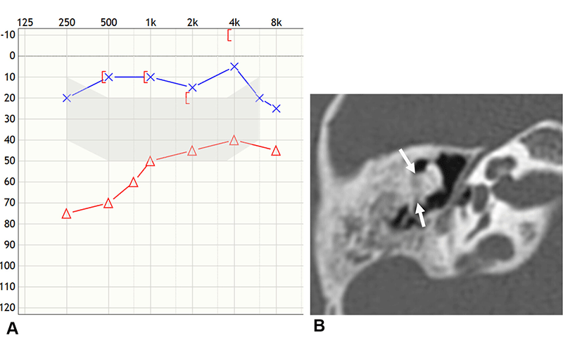
Tympanosclerosis in an 8-year-old boy who was evaluated for recurrent right ear infections. (A) Audiogram from pure-tone audiometry shows upsloping (ie, most pronounced at low frequencies) conductive hearing loss in the right ear. This audiogram configuration should prompt the radiologist to search for disease causing increased tympanic membrane or ossicular chain stiffness at temporal bone CT. (B) Axial temporal bone CT image shows abnormal mineralization (arrows) adjacent to the body and short process of the right incus, consistent with tympanosclerosis and appearing to fix the body and short process of the right incus to the lateral wall of the epitympanum. https://doi.org/10.1148/rg.240018 © RSNA 2024
Imaging and Audiologic Tests, All Pieces of the Puzzle
Temporal bone CT scans are a cornerstone of imaging in patients with conductive hearing loss. This imaging technique provides detailed views of the ear's intricate structures, including the ossicles, cochlea and semicircular canals. It can reveal abnormalities such as otosclerosis, cholesteatoma or congenital malformations that might not be apparent through clinical examination or audiologic testing alone.
Dr. Malouf commented that radiologists who are well-versed in both the technical aspects of these scans and the clinical context of hearing loss can make a significant difference in patient outcomes.
“I ended the research for this paper with a much better understanding of our ENT colleagues who see hearing loss, and a better appreciation for what we can bring to the table imaging wise to help sort out the treatment plan for patients,” he said.
For example, the clinical findings of third window lesions, which are bony defects that allow abnormal communication between the inner ear and the middle ear or cranial cavity, can mimic those of otosclerosis. Differentiation between the conditions is important because management is substantially different.
Audiologic tests provide valuable information about the type and severity of hearing loss, but imaging offers a different perspective that can be crucial in determining the underlying cause. Temporal bone CT is particularly useful in assessing conductive hearing loss, where sound waves are not efficiently conducted through the external and middle ear. By correlating audiologic findings with imaging results, radiologists can offer insights that might otherwise be missed.
“They are all different puzzle pieces that need to be assembled as a complete story,” Dr. Malouf said. “If you read the image in isolation, you may miss crucial pieces in different locations.”
A Collaborative Approach to Hearing Loss
Effective management of hearing loss often requires collaboration between various specialties, including otolaryngology, audiology and radiology. Dr. Malouf’s article highlights the importance of this multidisciplinary approach. By working closely with ENT specialists and audiologists, radiologists can help to ensure that all aspects of a patient’s hearing loss are considered in the diagnosis and treatment planning process.
For instance, in cases of conductive hearing loss, an ENT specialist might rely on the detailed imaging provided by a temporal bone CT to decide whether surgery is warranted. The radiologist’s ability to identify subtle abnormalities and correlate them with the patient’s clinical and audiologic profile is crucial in making these determinations.
The article makes the case for further integration of radiology into the broader framework of hearing loss management. As the population ages and the prevalence of hearing loss increases, the role of radiology in diagnosing and treating this condition will likely continue to expand.
Hearing loss is a complex condition that requires a nuanced and collaborative approach to diagnose and treat effectively. Dr. Malouf’s insights underscore the critical role that radiologists can play in this process, particularly using temporal bone CT imaging. By understanding the “puzzle pieces” of audiologic testing and imaging, radiologists can help put together the full picture of a patient’s hearing loss, leading to better outcomes and more personalized care.
“Optimal management commonly requires integration of clinical, audiologic and imaging information into a unifying diagnosis and individualized treatment plan,” the authors write. “Radiologists with a basic understanding of the relevant clinical and audiological evaluation of hearing loss are not only better positioned to effectively communicate with otologist and audiologist colleagues, but also to add value to the patient's care through confident and accurate image interpretation.”
For More Information
Access the RadioGraphics article, “Evaluation of Hearing Loss: Understanding Audiologic Testing to Refine Image Interpretation.”
Read previous RSNA News stories on head and neck imaging:
- Imaging Pulsatile Tinnitus: A State-of-the-Art Review
- What Radiologists Need to Know About Post-Treatment Head and Neck Imaging
- Sinonasal Tumors Receive New Classification From WHO