AI Screening Assistance Could Safely Alleviate a Third of Radiologists Mammography Workload
AI system implementation improved the cancer detection rate and decreased the false-positive rate

A population-based breast cancer screening study in Denmark suggests that AI could reduce the need for double readings of most mammograms—decreasing not only the workload for radiologists but also the rate of recalls and false positives.
“AI is not taking over the radiologists’ work,” said Andreas Lauritzen, PhD, from the Department of Computer Science at the University of Copenhagen, Denmark and Gentofte Hospital at the Capital Region of Denmark. “Rather, it assists and reallocates their time to more pressing tasks, freeing up time for them to focus on high-risk screenings or on diagnostic tasks for the patients who were identified for follow-up.”
Results Show Promise for Early Detection
In the screening program in the Capital Region of Denmark, a commercially available AI system was implemented in November 2021 to automatically select screening mammograms for single or double readings based on the probability that breast cancer is present. The AI system also aids radiologists by marking lesions in women at higher risk who are double-read.
The team’s retrospective analysis, published in Radiology, spanned multiple medical centers across the Capital Region of Denmark, including a “before AI” screening cohort of 60,751 individual women and a “with AI” cohort of 58,246.
Cases the AI selected for single reading encompassed about 70% of screenings, Dr. Lauritzen’s team found, reducing the workload for reading screening mammograms, by 33.5%.
And the findings came with a surprise benefit: better cancer detection overall. Detection before AI was 0.70% versus 0.82% after, and the false-positive rate decreased from 2.39% before AI to 1.63% with the AI system in place.
“We initially aimed to study AI for safely decreasing workload with non-inferior detection,” Dr. Lauritzen explained. “However, as a bonus, we observed that the cancer detection rate increased by 12 cancers per 10,000 screened women.”
“The size of invasive breast cancers decreased,” he added, “which could possibly indicate earlier detection.”

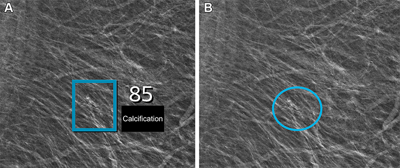
Left mediolateral oblique full-field digital mammographic view in a 67-year-old woman with a Breast Imaging Reporting and Data System density of 1 who underwent screening with the artificial intelligence (AI) system. (A) Image shows AI-provided marking (square). The screening received a high AI examination score of 10, based on this area with arterial calcifications being given a score of 85 out of 100 by the AI system. (B) Same image as in A, but with findings by the radiologists. Because of the high AI examination score, the screening was double read by two radiologists, who determined that the arterial calcifications (circle) did not yield suspicions for breast cancer. The woman was not recalled for diagnostic assessment.
https://doi.org/10.1148/radiol.232479 © RSNA 2024
Cautiously Skeptical Until Real-World Implementation
While the double-reading approach to screening mammograms improves accuracy, it adds substantially to the ever-growing workload and staffing issues that plague the specialty, wrote Amie Y. Lee, MD, and Sarah M. Friedewald, MD, in an accompanying editorial.
Dr. Lee is an associate professor of radiology who oversees breast imaging informatics at the University of California, San Francisco, and Dr. Friedewald is associate professor and chief of breast imaging at Northwestern University’s Feinberg School of Medicine in Chicago. They recalled the landmark study in the American Journal of Roentgenology (2006) by Constance Lehman, MD, PhD, and colleagues that demonstrated no improvement in accuracy from computer-aided detection (CAD), despite decades in clinical use.
“Although modern AI systems based on convolutional neural networks are superior to conventional CAD, repeating the same mistakes must be avoided and a dose of skepticism is warranted,” they cautioned.
This study from Dr. Lauritzen’s team answers the ever-amplifying call to move beyond the laboratory setting and to evaluate AI in a real-life screening environment, said Drs. Lee and Friedewald. It provides additional evidence that AI can help decrease workload while simultaneously preserving, if not increasing, the interpretive performance of screening mammography.
The authors noted some of the study’s limitations, which include the short follow-up period after screening (180 days) for biopsy and surgical outcomes. This minimum interval is too short to accurately assess false-negative findings, they asserted. And while the cancer detection rate was higher with AI assistance, the proportion of ductal carcinoma in situ was greater—as it was in the Mammography Screening With Artificial Intelligence (MASAI) trial in Sweden.
“Thus, the higher rate of cancer detection with AI support may not necessarily translate to the desired goal of reducing interval cancers and improving patient outcomes,” the editorial authors said.
And, naturally, screening settings differ in health care facilities across the world. European countries tend to have longer screening intervals and lower recall rates than health care centers in the U.S., for example.
Dr. Lauritzen and his team noted that results depend on the reading protocol, the radiologists and how the radiologists interact with the AI system.
“The takeaway,” Dr. Lauritzen said, “is that we can begin to trust that AI systems may be efficiently and safely implemented in breast cancer screening to reprioritize the radiologist’s workload.”
For More Information
Access the Radiology study, “Early Indicators of the Impact of Using AI in Mammography Screening for Breast Cancer.”
Read previous RSNA News articles on AI and mammography: