The Role of PET-MRI in Alzheimer's Disease and Dementia Care
Recently FDA-approved anti-amyloid monoclonal antibody therapy can potentially slow disease progression

RSNA News highlights the research being done in brain imaging to support Alzheimer's disease and dementia diagnosis and treatment. This is part four in a series of stories on this topic. Read part one, part two and part three.
PET-MRI offers a comprehensive approach to the evaluation and management of patients with Alzheimer’s disease (AD) and dementia, providing valuable insights into disease pathology, progression and treatment response. Its multi-modal imaging capabilities can enhance diagnostic accuracy and facilitate personalized patient care.
The recent FDA approvals of anti-amyloid monoclonal antibody therapies demonstrates the importance of PET-MRI to diagnose AD since this technology can simultaneously provide the required brain MRI for baseline safety as well as the required biomarker for beta-amyloid pathology.
“In order to be eligible for therapy, patients need a clinical diagnosis, confirmation of the amyloid pathology, and also a screening MRI to evaluate their risk for potential therapy complications, known as Amyloid Related Imaging Abnormalities (ARIA), which consist of brain edema (ARIA-E) and brain hemorrhages (ARIA-H),” said Tammie L. S. Benzinger, MD, PhD. “PET-MRI allows for completion of both aspects of this workup—MRI for safety and PET for amyloid confirmation— in a single visit, eliminating the burden on patients and families by requiring fewer medical appointments and improving the timeliness of the diagnostic workup. For a patient who has a positive amyloid PET on the PET-MRI, and no evidence of prior infarct or hemorrhage, the prognosis for therapy is very good.”
Dr. Benzinger is a professor of radiology, chief of MRI service and professor of neurological surgery, biology and biological sciences at Mallinckrodt Institute of Radiology at the Washington University School of Medicine in St. Louis.

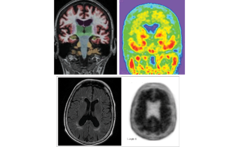
Fig Top. PET-MRI coronal brain T1 with quantitative structural segmentation for hippocampal volumes (left). Positive tau PET with 18F-flortaucipir (right).
Fig Bottom. PET-MRI axial brain FLAIR demonstrating mild white matter ischemic disease but no infarct (left). Positive amyloid PET with 18F-florbetapir (right).
Research images courtesy of Tammie L.S. Benzinger, MD, PhD
Benefits of Antibody Therapy for AD
For patients with early Alzheimer’s dementia, anti-amyloid monoclonal antibody therapies represent a significant milestone in AD research and drug development.
“Monoclonal antibody therapies have been shown in clinical trials to remove beta-amyloid from the brain on PET scans, and more importantly, to slow the clinical progression of early AD,” Dr. Benzinger said.
In the U.S., there is currently one approved anti-amyloid monoclonal antibody on the market, lecanemab. This therapy is now covered by the U.S. Centers for Medicare and Medicaid Services (CMS), and it is also covered by multiple private insurance carriers.
“For coverage approval, a clinical diagnosis of early AD, biomarker confirmation of abnormal beta-amyloid in the form of a PET scan or a lumbar puncture for CSF testing is required. More recently some insurance covers the use of a blood test for the latter,” Dr. Benzinger said. “A baseline MRI showing low risk of future ARIA, including lack of significant infarcts and hemorrhages at baseline, are the main findings that radiologists should look for. PET-MRI has a useful role in the patient workup as it can address two of the three required prerequisites for therapy in a single visit.”
These single visits for AD patients can be particularly effective for patients with cognitive impairment and their families, which can help with overall comprehensive care and inclusivity.
“PET MRI can help avoid diagnostic pitfalls and delays in care,” Dr. Benzinger said. “It also provides improved patient access to care because it eliminates multiple visits that are associated with reduced compliance for families of affected patients.”

Monitoring Response to Therapy
One of the ways that PET-MRI is used in patients with AD is in monitoring disease progression and response to therapy. Monitoring changes in amyloid burden over time with PET-MRI can provide insights into the effectiveness of anti-amyloid therapies.
PET-MRI also provides insight into brain glucose metabolism using tracers like 18F-FDG and assesses neuroinflammation in AD. Overall structural images of the brain allow for the assessment of brain atrophy, ventricular enlargement and other structural changes associated with AD progression.
Moving forward, PET-MRI will continue to play an important role in AD imaging.
“In addition to amyloid PET, diagnostic FDG PET and tau PET are also FDA-approved in the U.S. Many dementia centers complement structural MRI with perfusion MRI to evaluate the brain's perfusion, using either non-contrast arterial spin labeling (ASL) or gadolinium-enhanced techniques,” Dr. Benzinger said. “Research studies also employ MR diffusion sequences to evaluate neuro- inflammation, functional MRI for brain connectivity, and novel PET tracers to evaluate other pathologies, such as inflammation. Continued research and technological developments in this field are likely to further enhance the role of PET-MRI in imaging dementia and improving patient outcomes.”
For More Information
Read previous RSNA News stories on Alzheimer's disease research: