Upgraded PI-RADS 3 Prostate Lesions Are Less Likely to Harbor Clinically Significant Cancer
Study shows an upgrade to PI-RADS category 3 may result in unnecessary biopsies


New guidance for prostate cancer imaging could lead to more patients undergoing biopsies. Recognizing predictive features on multiparametric MRI could steer future guidelines to help ensure that those biopsies are not performed unnecessarily.
A 2019 update to the Prostate Imaging Reporting and Data System (PI-RADS), version 2.1, was released to further improve its reproducibility by clarifying the definitions of diffusion-weighted imaging and dynamic contrast-enhanced MRI scoring.
Version 2.1 also expanded its category 3 transition zone (TZ) to include nodules that exhibit marked diffusion restriction, meaning that a new subset of TZ lesions now meets the criteria for biopsy. But in a Radiology study, researchers for the National Cancer Institute (NCI) have found that the upgraded lesions were less likely to be cancerous than their non-upgraded counterparts.
“At many institutions, lesions that are categorized as PI-RADS category 3 or higher typically prompt a recommendation for biopsy,” explained the lead author Enis C. Yilmaz, MD, postdoctoral fellow at the NCI’s Molecular Imaging Branch. “PI-RADS version 2.1 introduced criteria allowing TZ lesions with a T2-weighted imaging score of 2 to ascend to category 3 if they exhibit diffusion-weighted scores of 4 or 5. However, our results demonstrated that upgraded category 3 TZ lesions were less likely to harbor clinically significant prostate cancer when compared to the nonupgraded category 3 TZ lesions, which may lead to unnecessary biopsy.”

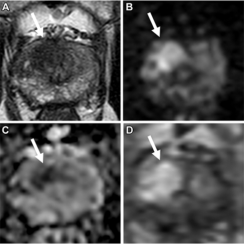
Multiparametric MRI in a 77-year-old man with an elevated prostate-specific antigen level of 10.7 ng/mL and a Prostate Imaging Reporting and Data System category 5 lesion located in the right apical-base anterior transition zone. (A) Axial T2-weighted image shows hypointense appearance of the lesion (arrow). (B) High–b value (b = 1500 sec/mm2) diffusion-weighted image shows high signal intensity of the lesion (arrow). (C) Apparent diffusion coefficient map generated using b values of 0, 300, and 600 sec/mm2 shows low signal intensity of the lesion (arrow). (D) Dynamic contrast-enhanced image shows early enhancement of the lesion (arrow). MRI/US fusion–guided biopsy revealed clinically significant prostate cancer with an International Society of Urological Pathology grade of 3. https://doi.org/10.1148/radiol.221309 ©RSNA 2023
Multiparametric MRI Improves Lesion Detection Based on Volume, Shape and Extraprostatic Features
Dr. Yilmaz and colleagues examined data from 454 men with suspected or known prostate cancer who underwent MRI/US fusion-guided biopsy. They obtained MRIs on a 3T scanner for each participant with T2-weighted imaging, diffusion-weighted imaging (including high b-value images and Apparent diffusion coefficient maps), and dynamic contrast-enhanced imaging sequences.
The investigators found that clinically significant prostate cancer rates differed notably among lesions in categories 5, 4 and 3—77%, 37%, and 14%, respectively.
“While there was no difference in clinically significant prostate cancer rates for category 4 TZ lesions, we have noted differences between nonupgraded and upgraded lesions for category 4 peripheral zone lesions and category 3 TZ lesions,” said principal investigator Baris Turkbey, MD, head of the Molecular Imaging Branch’s MRI and Artificial Intelligence Resource sections.
The difference amounted to a detection rate of just 4% in upgraded PI-RADS 3 transition zone lesions (i.e T2W score of 2 upgraded by the diffusion weighted score), versus 20% for nonupgraded PI-RADS 3 lesions (i.e. T2W score of 3).
“Considering these results, we may expect to observe an increased number of unnecessary biopsies for upgraded category 3 lesions,” Dr. Turkbey said. “Further prospective research is definitely needed to evaluate this.”
Imaging features on MRI like relative lesion volume, surface-to-volume ratio and extraprostatic extension scores of 2 and 3 were predictors of clinically significant prostate cancer, the investigative team found. Utilizing these factors could enhance the stratification of lesions, they said, suggesting that the next iteration of PI-RADS guidelines might benefit from considering these findings.
“Importantly, lesion PI-RADS categories and extraprostatic extension scores were also included in the multivariable analysis,” Dr. Yilmaz noted. “This means that the imaging features we identified are not driven by PI-RADS categories and extraprostatic extension scores and are independent predictors of harboring clinically significant prostate cancer.”
Current Iteration of PI-RADS Provides Appropriate Risk Stratification
In an accompanying editorial, Vicky Goh, MD, FRCR, a professor and chair of clinical cancer imaging at King’s College London, commented on the “balancing act” radiologists must perform to prevent overtreatment of indolent cancers while ensuring appropriate detection of clinically significant cancers.
“Given this trade-off of poorer specificity albeit with increased sensitivity, the jury remains out on the added value in substratification within PI-RADS 3 and PI-RADS 4,” Dr. Goh wrote. “There is an ongoing debate on whether the more objective assessment of shape and other radiomic and artificial intelligence approaches can improve the prediction of clinically significant prostate cancer.”
Until now, the lack of well-designed prospective studies and external validation has raised concerns about generalizability, noted Dr. Goh. She said that, despite limitations acknowledged by the authors, the study is an important step.
Both the authors and Dr. Goh touted the excellence of targeted biopsy versus systemic biopsy in MRI-defined lesions—in fact, the study by Dr. Yilmaz’s team excluded systematic biopsy results, including samples and reports from targeted biopsies only.
“Multiparametric MRI has a high negative predictive value, meaning that an individual with a negative MRI result is very unlikely to have the disease,” Dr. Goh explained. “A negative MRI finding can obviate unnecessary biopsy, and many prospective trials have demonstrated that an MRI-directed pathway is superior to a systematic ultrasound biopsy pathway in reducing the detection rate of clinically insignificant cancer and in confirming clinically significant disease.”
“Nevertheless,” she added, citing the multifocal nature of disease within the prostate, “clinically significant cancers can remain undetected in up to 26% of patients.”
Further Studies Necessary to Continue Identifying Markers for Prostate Cancer
Medical science is in a continual state of evolution, Dr. Yilmaz emphasized, and structured reporting systems like PI-RADS benefit from ongoing investigation and refinement.
“Envisioning a transition from subjective criteria to objective, reproducible, quantitative measures might be the key to addressing this challenge,” Dr. Yilmaz said. “Our study is an initial step towards this objective, and further research is imperative to continue this progression.”
For More Information
Access the Radiology study, “Prospective Evaluation of PI-RADS Version 2.1 for Prostate Cancer Detection and Investigation of Multiparametric MRI–derived Markers,” and the accompanying editorial, “PI-RADS and Multiparametric MRI: The Shape of Things to Come for Prostate Cancer.”
Read previous RSNA News stories on prostate cancer: