Top Images in Radiology Showcase Dramatic Views of Cutting-Edge Imaging Techniques
Unexpected presentations, striking appearances and unique views offer educational opportunities for radiologists
Selections have been made for the 2024 Images in Radiology, an online medical imaging collection that is part of the journal Radiology. Each year, Images in Radiology encompasses exceptional imaging representations of both common as well as extremely rare pathologies. It serves not only to highlight the significant contributions of medical imaging to clinical practice but also to provide an educational resource for radiologists and radiologists-in-training. This collection also demonstrates the striking examples of technological progress in radiology.
Images in Radiology is the second most popular manuscript submission type, after original research submissions. The Radiology In Training editorial board reviewed the Images in Radiology collection published from July 2023 through June 2024. Thirty eligible publications have been ranked on the basis of visual attractiveness, novelty of the imaging, techniques displayed, educational value and overall impact.
The top three images, second runner-up, first-runner-up and winner, were selected by the Radiology in Training editorial board based on aggregate scores from individual editorial board members’ rankings.

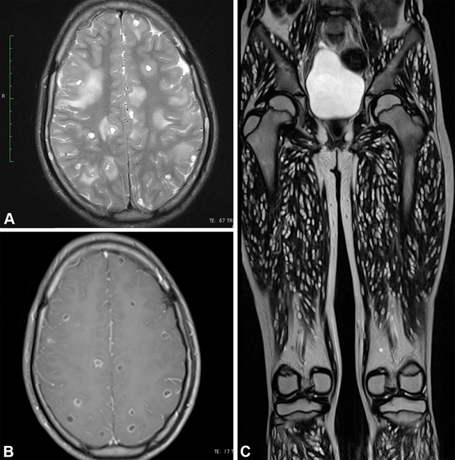
The 2024 Top Images in Radiology winner is entitled, “Disseminated Whole-Body Cysticercosis,” by Mohd Ilyas and Vikrant Mahajan. This case presents a 10-year-old female patient with generalized seizures, who underwent whole-body and brain MRI. Numerous cystic lesions in the brain, with ring enhancement and perilesional edema, together with a “rice grain” pattern of intramuscular cysts oriented parallel to muscle fibers, led to a diagnosis of disseminated cysticercosis. These striking images provoke a visceral reaction by revealing the body overwhelmed with parasites. At the same time, they teach readers the pathognomonic appearance of this entity, with multifocal small round cysts in the brain and oblong cysts in the muscles. This article was downloaded nearly 20,000 times and achieved an Altimetric score in the top 5% of all research outputs of the same age.

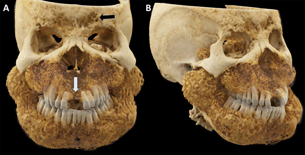
The first runner-up is the article entitled “Uremic Leontiasis Ossea,” by Dhairya A. Lakhani and Francis Deng. It highlights the case of a 34-year-old patient with end-stage renal disease and hyperparathyroidism who presented with facial hypertrophy for 6 months. Facial CT scan and subsequent cinematic volume renderings vividly demonstrated the marked maxillomandibular macrognathia, classically named “leontiasis ossea” due to the resulting resemblance with a lion’s face. Fibrous dysplasia, Paget disease, and chronic hemolytic anemia can mimic this appearance, but the serpentine intramedullary tunneling and cortical resorption in this case are characteristic of renal osteodystrophy.

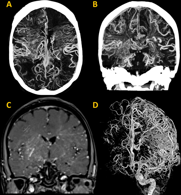
The second runner-up of 2024 Top Images in Radiology is “Diffuse Cerebral Proliferative Angiopathy,” by Dhairya A. Lakhani and SoHyun Boo. This article depicts the case of a 47-year-old woman presenting with transient episodes of left facial numbness, anomic aphasia, and left facial weakness. Head CT angiography and MRI revealed a diffuse network of vessels involving both cerebral hemispheres, with intermingled normal brain parenchyma. Angiography demonstrated diffuse pooling of contrast medium in the nidus with capillary ectasia, without dominant feeding, early draining vessels, or intracranial large-vessel stenosis. The final diagnosis was cerebral proliferative angiopathy, an extremely rare progressive vascular disease with fewer than 100 reported cases.
The other articles ranked as top 10 images in alphabetical order, included:
• Bone Marrow Edema at Photon-Counting CT (4)
• Brainstem Infarction Due to a Basilar Arterial Web (5)
• COVID-19 Hemiencephalitis: A Unique Manifestation (6)
• Diffuse Cavernous Hemangioma (7)
• Evolution of an Acute COVID-19 Pulmonary Infection (8)
• From Canvas to Screen: Resurrecting Artists of the Past (9)
• Interrupted Aortic Arch and Aortic Root Aneurysm and Dissection (10)
For More Information
Access the Radiology article, “2024 Top Images in Radiology: Radiology in Training Editors’ Choices.”
Review the 2023 Images in Radiology winners.
Review the 2022 Images in Radiology winners.
Review the 2021 Images in Radiology winners.