Putting CT Fractional Flow Reserve to the Real-World Test
CT-FFR shows potential to decrease rates of invasive angiography and coronary intervention


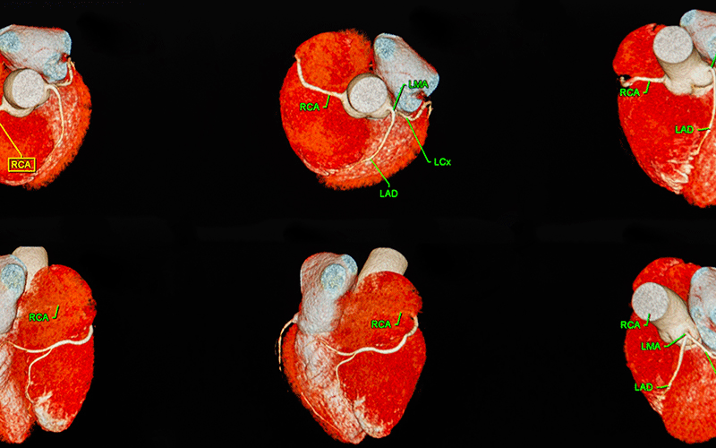
Coronary CT angiography (CCTA) is used to identify coronary artery disease and guide treatment decisions.
The challenge with CCTA is it cannot differentiate between those coronary stenoses that are physiologically significant from those that are not, despite their anatomic appearance.
“Identifying obstructions that are functionally insignificant can help safely defer invasive coronary angiography and revascularization,” said Mangun Randhawa, MD, a postdoctoral research fellow at Massachusetts General Hospital’s Division of Cardiovascular Imaging in Boston.
Making this distinction has traditionally involved using invasive physiologic measurements such as fractional flow reserve, a technique that measures blood pressure and flow in coronary arteries to determine the anatomical degree of a blockage.
But this is starting to change, with new noninvasive modalities like CT fractional flow reserve (CT-FFR) receiving approval for clinical use in the U.S.
“CT-FFR enhances the capabilities of CCTA by providing a noninvasive assessment of blood flow and pressure gradients across coronary stenoses, allowing us to better identify those lesions that necessitate invasive procedures,” said Brian Ghoshhajra, MD, MBA, MSCCT, division chief of cardiovascular imaging in the Department of Radiology at Massachusetts General Hospital and associate professor, Harvard Medical School.
While the addition of CT-FFR to CCTA has significantly improved pooled specificities compared to CCTA alone, most of the available data comes from vendor-sponsored trials.
But how does it perform in actual practice? That was the question posed by an article recently published in Radiology: Cardiothoracic Imaging.
CT-FFR Reduces the Need for Invasive Diagnostic Procedures
The retrospective observational study involved patients who underwent a clinically indicated CCTA scan during the large multisite practice’s initial roll-out of the test (2020–2021).
“Our analysis captured a number of clinical parameters, including age, gender, presence of hypertension, hyperlipidemia and baseline heart rates,” Dr. Ghoshhajra explained.
Researchers further examined CCTA features to assess coronary anatomy and the severity of stenoses, as well as the functional impact of the stenoses as predicted by CT-FFR. They also tracked clinical outcomes, including the use of invasive coronary angiography and percutaneous coronary intervention, the incidence of myocardial infarction and all-cause mortality.
In addition to being vendor agnostic, the study is unique in that its cohort was scanned without the use of heart rate control and that CT-FFR was applied selectively.
Researchers concluded that CT-FFR has the potential to reduce the need for invasive diagnostic procedures, particularly invasive coronary angiography. They also found the technique to be a reliable, non-invasive adjunct for assessing coronary artery disease.
“By reducing unnecessary procedures, the test reduces patient risk and health care costs, with the cost of the test being incurred at the discretion of the interpreting physician,” Dr. Randhawa said.
The study confirmed that the use of CT-FFR was most effective when triggered at the reading physician’s discretion than universally, which was the case in the sponsored trials.
This suggests that CT-FFR should be applied selectively to ensure the most efficient use of resources.
Improving Diagnostic Accuracy, Reducing Patient Risk
The study demonstrates a practical approach to integrating CT-FFR into clinical practice as a means of improving diagnostic accuracy, reducing patient risk and streamlining how coronary artery disease is managed.
“Radiologists can use the detailed insights into the functional impact of coronary lesions that CT-FFR offers to help physicians make more informed treatment decisions,” Dr. Randhawa concluded.
Further research is needed to assess the long-term outcomes of those patients managed using CT-FFR analysis, with performance evaluation undertaken via large-scale, multicenter studies to account for variations in both site and physician experiences.
For More Information
Access the Radiology: Cardiothoracic Imaging article, “Selective Use of CT Fractional Flow at a Large Academic Medical Center: Insights from Clinical Implementation after 1 Year of Practice.”
Read previous RSNA News articles about cardiac imaging:
- Low-income Country Cardiac Imaging Procedures Remain Down Post-COVID
- CT Test Simulates Blood Flow to Assess Risk in Patients with Angina
- Research Identifies Imaging Measurements and Serum Biomarkers Associated With Cardiac Strain and Myocardial Injury