AI Can Help Rule Out Abnormal Pathology on Chest X-Rays
Tool shows potential to deliver autonomous reporting with lower rate of clinically relevant errors than current standard

A commercial AI tool used off-label was effective at excluding pathology and had equal or lower rates of critical misses on chest X-ray than radiologists, according to a study published in Radiology.
Recent developments in AI have sparked a growing interest in computer-assisted diagnosis, partly motivated by the increasing workload faced by radiology departments, the global shortage of radiologists and the potential for burnout in the field. Radiology practices have a high volume of unremarkable chest X-rays, and AI could possibly improve workflow by providing an automatic report.
Researchers in Denmark set out to estimate the proportion of unremarkable chest X-rays where AI could correctly exclude pathology without increasing diagnostic errors. The study included radiology reports and data from 1,961 patients (median age, 72 years; 993 female), with one chest X-ray per patient, obtained from four Danish hospitals.
“Our group and others have previously shown that AI tools are capable of excluding pathology in chest X-rays with high confidence and thereby provide an autonomous normal report without a human in-the-loop,” said lead author Louis Lind Plesner, MD, from the Department of Radiology at Herlev and Gentofte Hospital in Copenhagen, Denmark. “Such AI algorithms miss very few abnormal chest radiographs. However, before our current study, we didn’t know what the appropriate threshold was for these models.”
The research team wanted to know whether the quality of mistakes made by AI and radiologists was different and if AI mistakes, on average, are objectively worse than human mistakes.
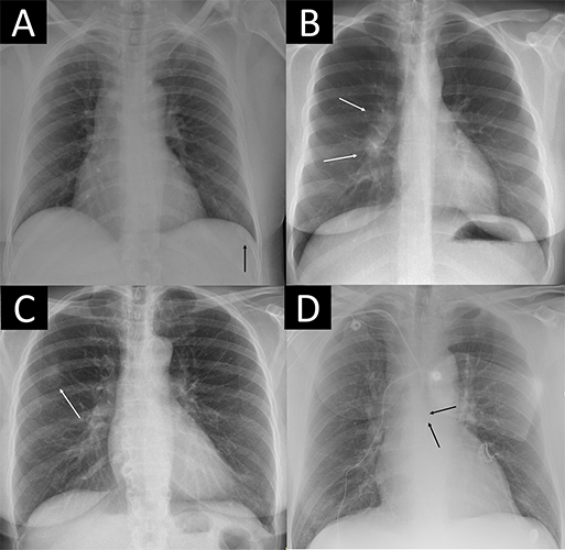
Four examples of remarkable chest X-rays with missed critical findings. The artificial intelligence (AI) tool was postprocessed by the AI vendor by scaling each of the 85 remarkable individual predictions to a normalized value and using the case-level highest of the scaled scores as the overall probability score from 0 to 1 for “remarkableness” (ie, the probability for abnormal or remarkable findings by the AI). (A) Chest X-ray in a 49-year-old female patient shows a slightly visible acute rib fracture (arrow) that was missed by the AI at all thresholds and also missed by the radiology report. (B) Chest X-ray in a 30-year-old female patient shows enlarged hilar lymph nodes (arrows) missed by the radiology report but not the AI at any threshold. (C) Chest X-ray in a 67-year-old female patient shows a tumor mimicking pleural plaque (arrow) that was reported in the radiology report (where the patient was referred for CT) and missed by the AI at the 98.0% threshold but not the 99.0% and 99.9% thresholds. (D) Chest X-ray in a 64-year-old male patient shows a central venous catheter possibly entering the azygos vein (arrows), which was classified as unremarkable in the radiology report. The AI missed the critical finding at the 98.0% threshold but not the 99.0% and 99.9% thresholds.
https://doi.org/10.1148/radiol.240272 © RSNA 2024
Additional Work Needed Before Widespread Deployment
The AI tool was adapted to generate a chest X-ray “remarkableness” probability, which was used to calculate specificity at different AI sensitivities.
Two chest radiologists, who were blinded to the AI output, labeled the chest X-rays as “remarkable” or “unremarkable” based on predefined unremarkable findings. Chest X-rays with missed findings by AI and/or the radiology report were graded by one chest radiologist—blinded to whether the mistake was made by AI or radiologist—as critical, clinically significant or clinically insignificant.
The reference standard labeled 1,231 of 1,961 chest X-rays (62.8%) as remarkable and 730 of 1,961 (37.2%) as unremarkable. The AI tool correctly excluded pathology in 24.5% to 52.7% of unremarkable chest X-rays at greater than or equal to 98% sensitivity, with lower rates of critical misses than found in the radiology reports associated with the images.
Dr. Plesner notes that the mistakes made by AI were, on average, more clinically severe for the patient than mistakes made by radiologists.
“This is likely because radiologists interpret findings based on the clinical scenario, which AI does not,” he said. “Therefore, when AI is intended to provide an automated normal report, it has to be more sensitive than the radiologist to avoid decreasing standard of care during implementation. This finding is also generally interesting in this era of AI capabilities covering multiple high-stakes environments not only limited to health care.”
AI could autonomously report more than half of all normal chest X-rays, according to Dr. Plesner. “In our hospital-based study population, this meant that more than 20% of all chest X-rays could have been potentially autonomously reported using this methodology, while keeping a lower rate of clinically relevant errors than the current standard,” he said.
Dr. Plesner noted that a prospective implementation of the model using one of the thresholds suggested in the study is needed before widespread deployment can be recommended.
For More Information
Access the Radiology study, “Using AI to Identify Unremarkable Chest Radiographs for Automatic Reporting,” and the related editorial, “Emerging AI Autonomy: Reducing the Burden of Unremarkable Examinations.”
Read previous RSNA News stories about the use of AI in radiography: