Radiologists Central to Burgeoning Field of Cancer Radiogenomics
Radiologists need to understand basic genomic concepts and their impact on imaging

Cancer radiogenomics combines imaging findings with genomic and epigenomic information in the search for new tumor classifications, biomarkers and targeted therapies. The field has developed rapidly in recent years.
“Because of technical advancements, we can do more next-generation sequencing, which allows us to sequence the genome to know the entire genetic composition of tumor cells,” said Xiaoyang Liu, MD, PhD, assistant professor, Division of Abdominal Imaging, Joint Department of Medical Imaging, University of Toronto. “The tumor’s genomic composition tells us if it’s going to be responsive to treatment or if it’s going to be very aggressive and should be treated differently.”
Cancer genomics is particularly well-suited for today’s push toward precision medicine, Dr. Liu commented. Combing radiology phenotypes with genomic information allows for non-invasive prediction of tumor subtype, tumor biological behavior and treatment response.
Because of this correlation of cancer genomics with imaging findings, radiologists need to be familiar with its basic concepts. Dr. Liu and colleagues outlined these concepts in a Radiology: Imaging Cancer article that reviewed links between genetic mutations and imaging findings in different cancers.

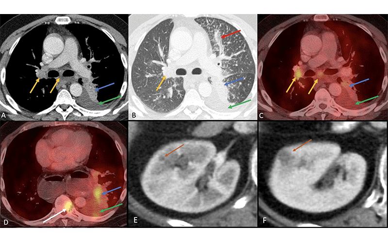
ALK mutation in non–small cell lung cancer. Axial images from (A, B) soft-tissue and lung window CT, (C, D) fusion PET/CT, and (E, F) postintravenous contrast CT of right kidney. Patient is a 53-year-old female who does not smoke, with non–small cell lung cancer with an ALK mutation who presented with (A–D) a central solid fluorodeoxyglucose-avid tumor (blue arrow) accompanied by pleural effusion (green arrow), metastatic mediastinal and hilar lymphadenopathy (yellow arrows), pulmonary lymphangitic carcinomatosis (red arrow in B) and vertebral bone metastasis (white arrow in D). Patients treated with crizotinib (ALK and ROS1 inhibitor) often develop new or increased renal cysts (orange arrow in E, F) with simple or complicated features as part of their treatment. These cysts often regress after stopping crizotinib. https://doi.org/10.1148/rycan.220153 ©RSNA 2023
Brain cancer
Research has identified several radiogenomic signatures of gliomas, the most common type of primary brain tumor, that can help guide treatment.
Patients with an IDH1/2 mutation, for instance, are more likely to benefit from targeted therapy. Imaging findings linked with these mutations include a larger tumor size with a distinct border, cortical involvement and less marked enhancement.
In contrast, patients with amplification and mutation of the epidermal growth factor receptor (EFGR) gene and a central or midline tumor location in the brain experience a poor prognosis and may require more aggressive treatment.
Lung cancer
Genomic characterization of lung cancer classifies tumors according to distinct mutations.
For example, EGFR mutations occur in approximately 15% of non-small cell lung cancer (NSCLC), a group of cancers that make up the vast majority of all lung cancers. Imaging features associated with EGFR mutations include pure or mixed ground-glass opacity, air bronchograms, pleural retraction, small size and the absence of fibrosis. After correlating genetic mutations with imaging features, clinicians can match patients with treatments that are far more targeted than conventional cytotoxic chemotherapies.
Breast cancer
Distinct imaging features such as tumor shape, margins, calcifications and size have been associated with different molecular subtypes of breast cancer. Estrogen receptor (ER) status and imaging features provide indications of treatment response.
For example, irregular margins and intratumoral necrosis correlate with a poor response to neoadjuvant chemotherapy. Peritumoral edema has also been related to reduced disease-free survival.
Gastrointestinal system
Hepatocellular carcinoma (HCC) represents about 90% of all primary liver cancers. The two most common mutations in HCC are in the TP53 and CTNNB1 genes. Imaging features for more aggressive tumors characterized by TP53 mutation include necrosis of more than 20% and arterial phase hyperenhancement. Features linked with less aggressive tumors with CTNNB1 mutations include persistent peritumoral hyperenhancement through arterial to delayed phase and well-defined margin.
Genitourinary system
A few well-recognized genetic mutations drive clear cell renal cell carcinoma, which accounts for 70% of kidney cancers. The presence of these mutations along with imaging features like ill-defined margins, calcifications and renal vein invasion are associated with more aggressive disease.
Bladder cancers that occur as a muscle-invasive urothelial carcinoma (MIUC) are linked with poor survival due to metastases. The Cancer Genome Atlas Research Network has reported 58 significantly mutated genes in MIUC. When reporting a scan in a patient with TP53 or RB1 mutations, the radiologist should be particularly attentive to equivocally enlarged or heterogeneous small lymph nodes, particularly in the pelvis, and search carefully for early signs of metastasis.
Understanding Genomics Positions Radiologists to Provide Patient-Centered Care
By incorporating these concepts into clinical practice, radiologists can make their imaging interpretations more meaningful and better facilitate multidisciplinary clinical dialogue and interventions, according to Dr. Liu.
“The care of cancer patients no longer involves a single expertise,” she said. “Radiologists, oncologists, surgeons, radiation oncologists and subspecialty medicine doctors must have a basic understanding of this field because we need to work as part of a team to improve patient care.”
Integration of radiology and genomics will become even more central to cancer care, Dr. Liu said, as results from prospective studies come in and access to tumor genomic sequencing expands. She expects the role of radiologists to expand along with it.
“Radiologists have a very pivotal role in cancer radiogenomics because we are at the intersection of imaging, diagnosis, treatment planning and interventional treatment,” Dr. Liu said. “We have the potential to be leaders in this field.”
For More Information
Access the Radiology: Imaging Cancer paper, “Current Status of Cancer Genomics and Imaging Phenotypes: What Radiologists Need to Know.”
Read previous RSNA News articles on various types of cancer: