Photon-Counting CT: A Game Changer in MSK Imaging?
Proponents point to benefits including vastly improved spatial resolutions


This is the third article in a series focusing on advances in MSK imaging. Read the first and second stories.
Musculoskeletal (MSK) imaging often deals with submillimeter structures best depicted using high-resolution imaging systems, such as computed tomography (CT).
With the introduction of photon-counting CT, radiologists can achieve unprecedented spatial resolution of individual trabeculae of cancellous bone. This is because photon-counting CT converts X-rays directly to electrical signals in a single step, rather than requiring two steps, as in conventional CT, according to Kishore Rajendran, PhD.
Dr. Rajendran is an assistant professor of radiology and research associate consultant at the Mayo Clinic’s CT Clinical Innovation Center in Rochester, MN. The institution was an early adopter of photon-counting CT.
“Unlike dual-energy CT, where we often face the dilemma of whether to acquire images at high spatial resolution or in dual-energy ‘spectral’ mode, we are now able to routinely acquire spectral data at high spatial resolution using photon-counting CT,” Dr. Rajendran said. “This streamlines our approach to protocol design and simplifies our workflow, as all MSK CT exams can be performed at high spatial resolution with spectral data always available.”
Dr. Rajendran was the senior author on the subject in an American Journal of Roentgenology article offering a clinical perspective on the use of photon-counting CT in MSK imaging.
His co-author, Cynthia H. McCollough, PhD, professor of medical physics and biomedical engineering and director of the Mayo Clinic’s CT Clinical Innovation Center, said photon-counting CT measures—or counts—each individual X-ray and its energy. This is something conventional detectors cannot do, she noted.
“The benefits of photon-counting detectors include improved spatial resolution, reduced image noise, increased iodine or calcium signal and the availability of multi-energy information in all scan modes,” she said. “Studies have shown not only improved image quality but also reduced radiation dose.”
Photon-counting CT improves the quality of CT for all applications, but is particularly beneficial in MSK, cardiac, vascular, lung and inner-ear imaging, Dr. McCollough said.
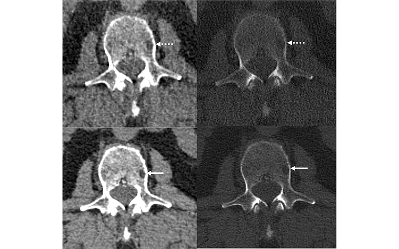
Francis Baffour, MD, associate medical director of the Mayo Clinic CT Clinical Innovation Center and lead author of a Radiology study on photon- counting CT for multiple myeloma, noted that higher energy X-rays have greater potential to reduce metal artifact.
“With photon-counting CT detectors, high-energy threshold images can be leveraged to produce images with reduced metal artifacts and beam hardening effects,” he said.
In the Radiology study, he and his fellow researchers found that imaging features of myeloma were depicted more clearly on photon-counting detector images compared with energy-integrating detector images.
“We believe that the lower image noise and higher intrinsic spatial resolution of the photon-counting detector images led to sharper lesion margins and better image contrast,” he said. “In phase two of the study (with photon- counting detector images at thinner image sections of 0.6 mm and deep learning denoising), the radiologists detected more lytic lesions compared to energy-integrating scans.”
The Future of Photon-Counting CT
The potential applications of photon-counting for MSK imaging may include trauma, rheumatology, osteoporosis, oncology and pre- and post-operative settings, according to Patrick Omoumi, MD, PhD, head of musculoskeletal imaging at Lausanne University Hospital and the University of Lausanne in Switzerland.
Dr. Omoumi is co-author of a Skeletal Radiology study that explored the technical advantages of photon-counting CT.
“The improved spatial resolution is particularly useful for the imaging of trabecular and cortical bone, aiding in the detection of subtle fracture and callus formation,” he said. “Furthermore, the high resolution of photon-counting CT could provide qualitative and quantitative assessment of bone architecture on clinical scanners, which is usually reserved for high resolution peripheral quantitative CT (HR-pQCT) but is limited to the extremities. This could have applications for the diagnosis of conditions such as osteoporosis.”
Dr. Omoumi added that “the improved resolution of photon-counting CT may enhance the detection and quantification of crystal deposits.”
In addition, the lower radiation dose may extend the use of CT for certain indications such as 4D CT for chronic joint instability, or low-dose whole-body CT for the detection of skeletal metastases.
“The enhanced spectral capabilities have the potential to improve the detection of bone marrow edema—for the detection of occult fractures, for instance—and decrease metal artifacts,” Dr. Omoumi said. “The differentiation between types of crystal deposits in those arthropathies may be improved. Finally, new applications such as multicontrast compositional imaging of joint tissues may emerge.”
Managing Challenges
As with any new technology, Dr. Rajendran noted, there is a learning curve associated with tailoring photon-counting CT protocols to meet the needs of an imaging practice.
“With the high spatial resolution produced by photon-counting CT, users can anticipate the image number and matrix size to increase compared to older CT technologies,” he said. “Therefore, sites should assess whether their existing PACS solutions can accommodate the types and volume of image data produced by photon-counting CT.”
While this technology has great potential for current applications of CT, as well as for new and emerging applications, thorough clinical evaluation is still underway to show clinical benefit and cost-effectiveness, Dr. Omoumi noted.
“The theoretical advantages of photon- counting detector CT in terms of improved diagnostic performance still need validation on a larger clinical scale,” he said
For More Information
Access the Radiology study, “Photon-counting Detector CT with Deep Learning Noise Reduction to Detect Multiple Myeloma."
Access the American Journal of Roentgenology article at AJR.org
Access the Skeletal Radiology paper at Link.Springer.com/Journal/256.
Read previous RSNA News articles on MSK imaging: