Latest Lung-RADS Version Improves Specificity of Cancer Detection
The 2022 update reduced the number of false-positive screening CT exams

The latest version of a widely used system for classifying lung nodules reduces the number of false-positive screening CT examinations without sacrificing sensitivity for cancer detection among airway nodules, according to research from Radiology: Cardiothoracic Imaging.
Annual lung cancer screening with low-dose CT is recommended for people at high risk of lung cancer such as those with a history of heavy smoking. The 2022 update to the Lung Imaging Reporting and Data System (Lung-RADS) included revised assessment criteria for airway nodules, also known as endobronchial nodules. Version 1.1, which was updated in 2019, classified all endobronchial nodules as suspicious lesions (category 4A). In the new version, airway nodules at baseline are classified as either benign (category 2) or category 4A based on imaging features.
“Previously, all airway findings were classified as category 4A,” said study lead author Ariadne DeSimone, MD, MPH, a thoracic radiologist at Brigham and Women’s Hospital (BWH) in Boston. “In our study when applying the updated classification system, many of the airway nodules could be reclassified as benign.”

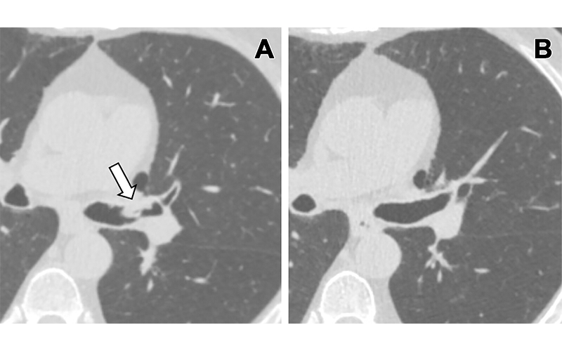
(A) Lung cancer screening CT image (cropped axial plane image, without contrast agent) in a 66-year-old male patient shows a sessile nodule with internal air in the left mainstem to left upper lobe bronchus (arrow) with a mean diameter of 10 mm. The nodule was assigned as Lung Imaging Reporting and Data System category 4A in the clinical report. (B) Follow-up CT image shows the lesion is resolved. https://doi.org/10.1148/ryct.230149 © RSNA 2024
Updated Version Helps Reduce Unnecessary Tests
For the study, radiologists at BWH analyzed the effects of the updated Lung-RADS on patients who underwent a lung cancer screening CT examination with a reported airway or endobronchial nodule.
Of the 174 patients, 163 (94%) had airway nodules that were deemed benign, while 11 (6%) had malignant nodules. Lung-RADS version 2022 maintained the 91% sensitivity for cancer detection demonstrated with version 1.1 while delivering a much-improved specificity of 82% compared with only 50% for the previous version. The researchers attributed the improved specificity to a reduction in false positive results.
The improved classification system is important because further workup of 4A nodules may require a bronchoscopy with biopsy or percutaneous lung biopsy.
“Both procedures have risks associated with them,” Dr. DeSimone said. “Reducing false-positive studies means reducing potential unnecessary interventions.”
Better At Characterizing Lesions
The findings support previous research that shows the vast majority of airway nodules or endobronchial lesions are benign. Nodules located in the trachea and mainstem bronchi in the study group were consistently benign. Multiple airway nodules were also more likely to be benign.
Benign features, such as nonobstructive morphologies, dependent portion of the airway, internal air, or fluid attenuation further indicated a lower risk of malignancy. In two previous studies, some of these imaging features were used to differentiate between mucus secretions and airway tumors on CT.
“Lung-RADS version 2022 was a useful update taking into account various imaging features such as location, morphology, number, and stability or growth to better characterize the airway or endobronchial lesions,” Dr. DeSimone said. “However, there is some remaining ambiguity if there is an obstructive airway nodule at the subsegmental level.”
The subsegmental bronchi are the very small branches of the airways located in the more distal part of the tracheobronchial tree at the periphery of the lungs. Lung-RADS v2022 categorizes nonobstructive subsegmental airway nodules as category 2, but it does not specify a category for obstructive subsegmental airway nodules.
In the study, more than a quarter of the malignancies occurred in these subsegmental bronchi, indicating that subsegmental location of an airway nodule does not guarantee that a nodule is benign. Notably, of the subsegmental airway nodules that were obstructive in the study, 25% were malignant.
“We would recommend that obstructive airway nodules at any level be upgraded to at least Lung-RADS category 4A,” Dr. DeSimone said.
Despite the encouraging findings, the researchers recommend that future updates address certain obstructive airway lesions that have a significantly increased risk of malignancy.
“Radiologists must remain wary of obstructive endobronchial lesions at any airway level because these have a significantly increased risk of malignancy,” Dr. DeSimone recommended. “Future studies are warranted to confirm these findings in larger cohorts.”
Editor’s Note: Dr. DeSimone’s BWH colleagues Suzanne Byrne, MD, and Mark Hammer, MD, coauthored the study.
For More Information
Access the Radiology: Cardiothoracic Imaging study, “Comparison of Lung-RADS Version 1.1 and Lung-RADS Version 2022 in Classifying Airway Nodules Detected at Lung Cancer Screening CT.”
Read previous RSNA News stories on lung cancer: