The Potential of Automated Opportunistic Imaging to Combat Sarcopenia's Stealth
Radiology provides innovative data gathering and reporting to assist with early detection and prevention of sarcopenia


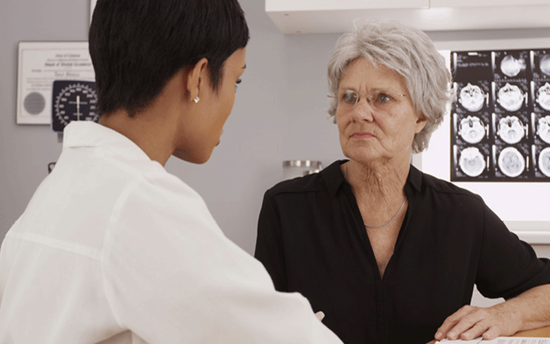
RSNA News highlights the role radiology plays in supporting the imaging needs of the aging population. This is the first in a series of stories. Read the second and third story.
Sarcopenia, the age-related, progressive loss of muscle mass and strength, is often called a “hidden” disease. It is usually an incidental finding of diagnostic tests for other diseases and is not routinely screened for.
Despite being hidden, the effects of sarcopenia are very apparent: increased risk of falls and related injuries, longer hospital stays, loss of function, worse surgical outcomes, and even death.
“Sarcopenia affects up to 10% of community-dwelling elderly patients and approximately 20% of hospitalized patients, and it is associated with increased risk of falls, longer hospital stays, postoperative complications, and mortality,” said Jennifer Padwal, MD, MAS, radiology resident at Stanford Medicine in California.
Luca Maria Sconfienza, MD, PhD, professor of radiology at University of Milano and chair of the Radiology Department at IRCCS Galeazzi-Sant’Ambrogio Research Hospital, Milano, Italy, pointed out that good muscular health is vital for the mobility and independence of older adults.
“Many people think that a good muscle mass is needed only for athletes; this is totally wrong.” he said. “If osteoporosis is related to bone fragility, muscles are those organs which keep the skeleton standing or enable motion: if muscles fail, the patient falls and may fracture bones. Further, reduced muscle mass implies reduced mobility, which in turn results in loss of self-sufficiency.”
As the population ages worldwide, the prevalence of sarcopenia is expected to rise: 50 million people were diagnosed with sarcopenia in 2010, and in 2050, that number is projected to reach 250 million. Radiology can play a role in early diagnosis and prevention through opportunistic imaging and automated reporting.
Providing Imaging Without Additional Acquisition Time or Cost
Ideally, sarcopenia is diagnosed through clinical assessments for muscle strength, followed by imaging studies as confirmation, as set out by the European Working Group on Sarcopenia in Older People guidelines.
However, because sarcopenia is not routinely screened for, it is most often detected secondarily through opportunistic imaging.
“Opportunistic imaging, or the additional use of existing imaging to obtain clinical information separate from the primary indication, has the potential to help identify sarcopenic patients and facilitate early intervention without incurring additional cost, radiation, or acquisition time,” Dr. Padwal said.
Dr. Padwal added that ongoing opportunistic imaging can also provide longitudinal monitoring of sarcopenia treatment response.
Although sarcopenia can be assessed on all imaging modalities besides conventional radiography, CT and MRI provide rich qualitative and quantitative data on sarcopenia through cross-sectional imaging.
“CT and MRI have the great advantage of providing cross-sectional imaging, very high accuracy and reproducibility,” Dr. Sconfienza said. “In both modalities, images of sarcopenic patients present with muscles with increased fat content, or myosteatosis, and reduced cross-sectional area. Important parameters are related to numerical assessment of images, such as muscle density on CT or muscle fat fraction on MRI.”
Where sarcopenia is the primary target of an imaging study, dual-energy X-ray absorptiometry (DXA) is the modality of choice.
“Whole-body DXA should be the preferred modality when sarcopenia is assessed primarily,” Dr. Sconfienza explained. “It allows concurrent measurement of lean mass, fat mass, and bone mineral content.”
Automated Sarcopenia Assessment Possible
Given that sarcopenia is visible on many imaging modalities, routine assessment of sarcopenia, through opportunistic imaging—with the help of machine learning—presents an opportunity for early detection and prevention.
Lawrence Yao, MD, research consultant in radiology and imaging sciences at the National Institutes of Health, Bethesda, MD, sees a near future wherein sarcopenia reporting is automated and included in reports for other common, diagnostic tests.
“Sarcopenia reporting as part of other diagnostic exams is already happening, but in order to be more widespread, it’s a matter of agreeing on what the standard measures are going to be,” Dr. Yao said. “There’s a lot of people working on standardized metrics for sarcopenia on imaging studies that can be automated using machine learning quite reproducibly.”
Dr. Sconfienza noted that sarcopenia assessment can be time consuming for radiologists and may be best left to AI in order to reduce radiologists’ workloads.
“Correctly assessing sarcopenia on a CT scan requires specific competence and additional time, which may not be available in clinical practice,” Dr. Sconfienza said. “Having an AI program that automatically segments muscles and calculates sarcopenia parameters in these examinations could be of great added value. This could be highlighted in the radiologic report as a kind of ‘side information,’ which then should be worked up in a specific clinical setting.”
The possibility of automating sarcopenia reporting through deep learning on routine imaging exams could lead to a shift in the way that health care professionals think of sarcopenia. Rather than a condition that commonly afflicts older people and can’t be avoided, it may become something that can be detected early, and delayed or reversed through interventions.
“It would be beneficial to address these things throughout the patient’s lifespan, rather than waiting until the point where it’s much harder to reverse some of these processes,” Dr. Yao said. “Now that people are understanding the implications of sarcopenia, there’s a lot more interest in prevention, through things like resistance training and nutrition.”
For More Information
Access the Abdominal Radiology study, “Sarcopenia: imaging assessment and clinical application,” at Link.Springer.com/Journal/261.
Access the Skeletal Radiology study, “Diagnosing sarcopenia at the point of imaging care: analysis of clinical, functional, and opportunistic CT metrics,” at Link.Springer.com/Journal/256.