CT Health Screening Can Identify Diabetes Risk
Automated CT analysis improves risk prediction and early intervention strategies

Analysis of CT scans in people who undergo imaging for health screening can identify individuals at risk of Type 2 diabetes, according to a study published in Radiology. Researchers said the findings underscore CT’s value in opportunistic imaging.
For the new study, researchers evaluated the ability of automated CT-derived markers to predict diabetes and associated conditions.
“Given the significant burden of diabetes and its complications, we aimed to explore whether automated and precise imaging analyses could enhance early detection and risk stratification beyond conventional methods,” said study senior author Seungho Ryu, MD, PhD, from the Kangbuk Samsung Hospital at Sungkyunkwan University School of Medicine in Seoul, South Korea.
The study group included 32,166 adults ages 25 years or older who underwent health screening with 18F-fluorodeoxyglucose (18F-FDG) PET/CT.
Dr. Ryu and colleagues used clinically validated deep learning algorithms to analyze the CT images. The algorithms enabled 3D segmentation and quantification of various body components such as visceral fat, subcutaneous fat, muscle mass, liver density and aortic calcium.
Diabetes prevalence was 6% at baseline and incidence was 9% during the 7.3-year median follow-up.
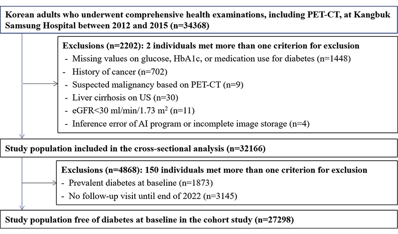
Flowchart of the selection of the study participants. AI = artificial intelligence, eGFR = estimated glomerular filtration rate, HbA1c = glycated hemoglobin.
https://doi.org/10.1148/radiol.233410 © RSNA 2024
Expanding the Role of CT Imaging
Automated multiorgan CT analysis identified individuals at high risk of diabetes and associated conditions. The index of visceral fat showed the highest predictive performance for diabetes.
Combining visceral fat, muscle area, liver fat fraction and aortic calcification improved predictive performance. CT-derived markers also identified ultrasound-diagnosed fatty liver, coronary artery calcium scores of more than 100, osteoporosis and age-related muscle loss.
These markers outperformed traditional risk factors in predicting Type 2 diabetes.
“The results are encouraging as they demonstrate the potential of expanding the role of CT imaging from conventional disease diagnosis to opportunistic proactive screening,” Dr. Ryu said. “This automated CT analysis improves risk prediction and early intervention strategies for diabetes and related health issues.”
In the clinical setting, these CT-derived markers have the potential to improve the conventional approach to diabetes screening and risk assessment, Dr. Ryu noted.
“By integrating these advanced imaging techniques into opportunistic health screenings, clinicians can identify individuals at high risk for diabetes and its complications more accurately and earlier than the current approach,” he said. “This could lead to more personalized and timely interventions, ultimately improving patient outcomes.”
For More Information
Access the Radiology article, “Automated Comprehensive CT Assessment of the Risk of Diabetes and Associated Cardiometabolic Conditions,” and the related editorial, “Harnessing the Value of Incidental Tissue and Organ Data at Body CT.”
Read previous RSNA News stories about CT imaging: