Understanding How Pregnancy and Breastfeeding Impact Breast Imaging and Diagnosis
Radiologists can help identify appropriate imaging for patients


In concert with the RadioGraphics monograph, RSNA News shares the latest in breast cancer research, intervention and patient education.
Women who are pregnant or breastfeeding experience physical changes that may make breast cancer screening and diagnosis more challenging, but that ought not delay diagnostic imaging.
For women who are pregnant or lactating, hormones can change breast density and size, which could limit the clinical examination, mimic pathology and obscure mammographic findings.
Pregnancy-associated breast cancer is routinely defined as breast cancer that occurs during pregnancy or within one year postpartum. Breast cancer is the most common type of invasive cancer diagnosed in pregnant and postpartum women with an incidence of one in 3,000 in the U.S. This incidence is increasing as more women postpone childbearing until later ages, according to a RadioGraphics education review.
“Our goal was to draw attention to the importance of this issue and make sure that women are worked up appropriately during and after pregnancy,” said Molly Peterson, MD, lead suthor on the article and a breast imaging fellow at the University of Wisconsin-Madison. “Pregnancy- associated breast cancer is rare, but it is associated with worse outcomes and so we want to make sure these women feel comfortable with imaging and that physicians who see them are not dismissing any symptoms.”

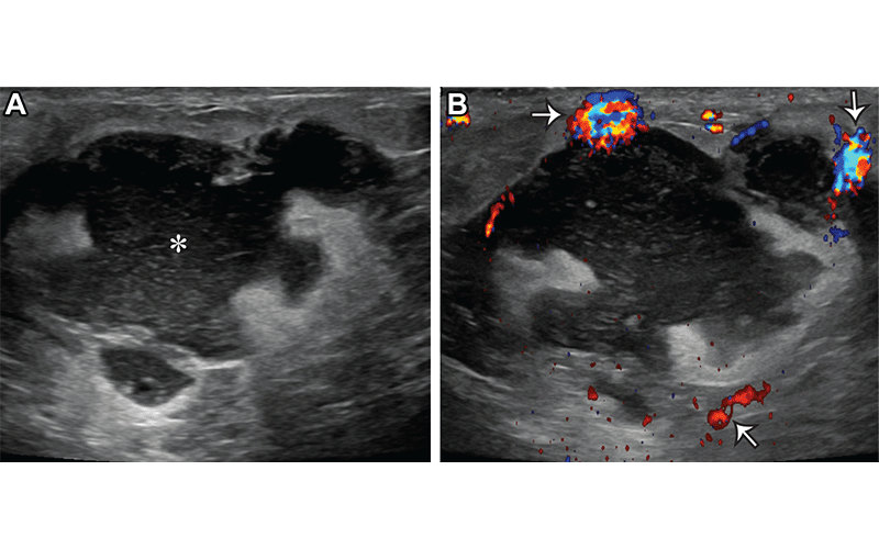
Acute puerperal abscess in a 26-year-old lactating woman who presented with skin erythema, warmth, and pain. (A) Gray-scale US image shows an irregular complicated fluid collection (*). (B) Color Doppler US image shows hyperemia surrounding the fluid collection (arrows). US-guided aspiration yielded purulent fluid, and cultures grew methicillin-resistant Staphylococcus aureus (MRSA). https://pubs.rsna.org/doi/10.1148/rg.230014 ©RSNA 2023
How Pregnancy and Breastfeeding Affect Imaging
During pregnancy, the breasts undergo physiologic changes in response to hormones to prepare for lactation. Women’s elevated estrogen and progesterone levels can lead to enlarged ducts and lobules, as well as increased breast density that can make imaging and diagnosis more challenging.
“It’s important to recognize imaging findings that are clearly benign that might be associated with pregnancy and breastfeeding versus those that are suspicious and need to be biopsied,” said Amy Fowler, MD, PhD, associate professor of radiology in the breast imaging section at the University of Wisconsin-Madison. “For women, if there is an area that feels different from the rest of the breast, even though the whole breast feels lumpy from breastfeeding, be sure to let a doctor know and they can order a mammogram and ultrasound to further evaluate.”
88% of pregnant, lactating and postpartum women presenting for breast imaging evaluation have dense or extremely dense breasts. Breastfeeding or pumping before imaging may be helpful, according to Dr. Fowler. Breast abnormalities most often have a benign cause, such as an abscess, galactocele or mastitis, but in some cases may be something more serious and women should not postpone diagnostic imaging.
“It’s important to recognize things that are clearly benign that might be associated with pregnancy and breastfeeding versus things that are suspicious and need to go on for biopsies. For women, if there is an area that feels different from the rest of the breast, even though the whole breast feels lumpy from breastfeeding, be sure to let a doctor know and they can order a mammogram and ultrasound to further evaluate.”
AMY FOWLER, MD, PHD
Breast Imaging Safe and Sensitive for Pregnant and Lactating Women
The American College of Radiology (ACR) Appropriateness Criteria emphasize that mammography for both screening and diagnostic indications is safe during pregnancy and lactation for both screening and diagnostic indications. For a fourview mammogram examination, the fetal radiation dose is less than 0.03 mGy, a negligible amount, according to the article.
“In the situation where there is a lump or a specific finding that needs to be evaluated, the importance of promptly diagnosing breast cancer outweighs the very tiny risk of radiation to the fetus,” Dr. Fowler said.
RSNA also has a position statement on imaging safety during pregnancy, which recognizes the important role a radiologist plays in identifying the appropriate imaging test during pregnancy or lactation. There may also be questions about test sensitivity during a time of so much physical change, but that is not the case.
“Studies show that mammography is actually still quite sensitive for detecting breast cancer in that patient population,” Dr. Peterson said.
Despite increased breast density, the sensitivity of mammography for detecting breast cancer has been reported to range from 74% to 100% during pregnancy and lactation. US is the preferred initial imaging modality during pregnancy as it has greater sensitivity than mammography and confers no radiation exposure to the fetus and expectant mother. Most studies report the sensitivity of US for the detection of pregnancy-associated breast cancers as nearly 100%.
In addition, breast MRI can be safely performed for lactating women, but it is not recommended during pregnancy. Changes to the breast related to lactation were initially hypothesized to limit the sensitivity of MRI for breast cancer detection, however several studies have shown that the sensitivity of breast MRI for detecting breast cancer during lactation is 98% to 100%.
The RadioGraphics article includes several imaging examples to help radiologists learn about the different presentations of breast-related issues in pregnant and lactating women and serve as a guide for radiologists working with this sensitive population, which is why Dr. Fowler said this topic is so important to her.
“It is important for radiologists to know the appropriate imaging evaluation and management in order to promptly diagnose pregnancy-associated breast cancer and correctly distinguish it from more common benign findings,” Dr. Fowler concluded.
For More Information
Access the RadioGraphics paper, "Breast Imaging and Intervention during Pregnancy and Lactation."
Read previous RSNA News stories on breast cancer imaging: