Gantry-Free Cone-Beam CT Offers More Flexibility for Elbow Imaging
Patients who are unable to position their elbow can benefit from quicker, clearer imaging

While elbow injuries are a common sight in the emergency room, quality images of those injuries can be difficult to achieve.
“Cross-sectional elbow imaging in the context of trauma can be quite challenging,” according to Andreas Kunz, MD, a radiologist at the University Hospital Würzburg in Germany.
According to Dr. Kunz, most CT systems have an opening diameter (the gantry) of 70 to 80 cm, meaning the optimal positioning for this type of injury requires patients to lie prone and extend the injured arm in a “superman” stance.
“The problem is that, whether due to a limited range of motion because of a cast or pain, many patients are simply unable to extend their arm forward,” he said.
Instead, the injured arm must be positioned beside the patient or on the abdomen, both of which negatively impact image quality and require a substantial increase in radiation dose.

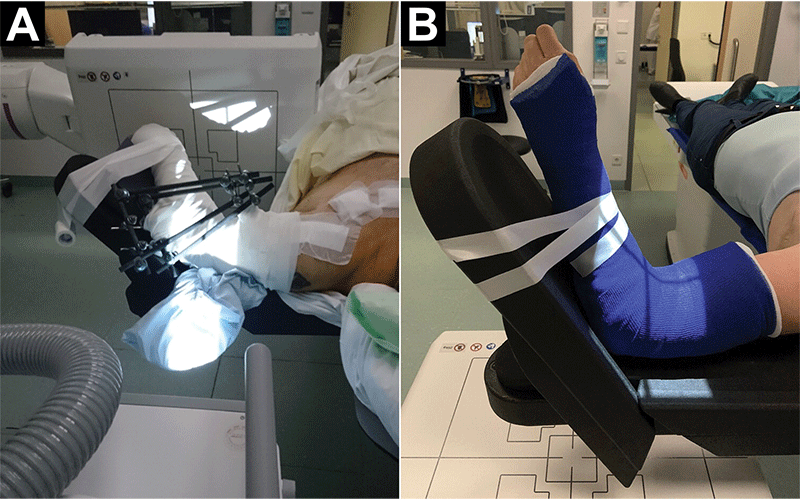
Photographs show patients with severely limited range of motion. In patients with (A) external fixation for concomitant shoulder injuries or (B) cast immobilization, the gantry-free system facilitates elbow cone-beam CT imaging with high spatial resolution in a comfortable tableside scan position. https://doi.org/10.1148/radiol.221200 ©RSNA 2022
Gantry-Free CBCT vs. 2D Radiography
The combined use of gantry-free cone-beam CT (CBCT) and large flat-panel detectors has emerged as an effective alternative for imaging the appendicular skeleton. In fact, studies have shown that gantry-free CBCT can offer a substantial dose reduction over gantry-based multidetector CT (MDCT) without penalizing image quality.
But what about its use with acute elbow trauma?
To answer this question, Dr. Kunz led a study on the diagnostic performance of gantry-free CBCT versus two-dimensional radiography in adults and children with acute elbow trauma.
The study, published in Radiology, used an ultra-high resolution CBCT scan mode based on a radiography system with two telescopic arms mounted on ceiling rails (see image below). One arm carries the X-ray tube, while the other holds a large flat panel detector. Simultaneous arm movement along predefined trajectories allows for three-dimensional projection data acquisition.
“The system’s use of a comfortable tableside armrest means the elbow can be placed in a flexed position at 90° abduction, essentially eliminating joint mobility or cast fixation as a limiting factor,” Dr. Kunz said.
High-Quality Images, Lower Dosage
In the retrospective study, elbow fractures in 23 patients with positioning difficulty in gantry-based CT were diagnosed with higher sensitivity using the gantry-free CBCT than what was achieved using traditional two-dimensional radiography.
“Not only did this CBCT system eliminate the positioning challenges associated with gantry-based CT, it also provided remarkably high-quality images,” Dr. Kunz noted.
In addition, the gantry-free CBCT offered superior dose efficiency, the result of the twin robotic system’s unique acquisition geometry.
“Instead of symmetric intervals between tube and patient and patient and detector, the two telescopic arms facilitate an asymmetric source-to-image distance with low magnification,” Dr. Kunz explained.
One concern Dr. Kunz had was that the CBCT system’s longer acquisition time (14 seconds compared to the five second scan time of a gantry CT) could result in increased motion artifacts and inferior image quality. However, because the system allows the elbow to be scanned in a comfortable, flexed 90° position, patient movement was not an issue.
The Freedom of Positioning
Allowing for a much larger degree of freedom in patient positioning, the University Hospital Würzburg is now using gantry-free cone-beam CT systems instead of conventional gantry-based systems on a regular basis—not only for the elbow, but also for the wrist, hand and, in some cases, the foot.
In a related study, Dr. Kunz also demonstrated that imaging of the appendicular skeleton using the gantry-free CBCT system in the presence of potentially image-impairing metal implants is feasible.
“The investigated gantry-free cone-beam CT promises to alleviate the most common challenges to imaging the appendicular skeleton without compromising image quality, radiation dose or patient comfort,” Dr. Kunz said.
For More Information
Access the Radiology study, “Twin Robotic Gantry-Free Cone-Beam CT in Acute Elbow Trauma.”
Read previous RSNA News stories on MSK imaging:
- Bone Tumor Identification Can Benefit from Deep Learning Model
- AI Helps Radiologists Detect Bone Fractures
- Cryoablation Effective in Relieving Pain in Bone Metastasis