Intranasal Drug Administration May Overcome the Blood-Brain Barrier Limitations
R&E grant investigates an alternative to systemic administration


Delivering therapeutic drugs into the brain through the almost-impenetrable blood-brain barrier is a significant ongoing challenge.
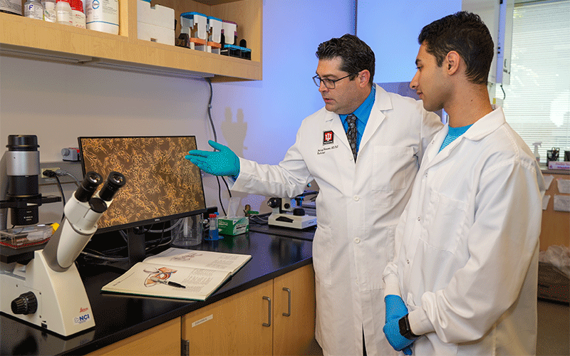
“Researchers are looking for novel ways that might be the Trojan horse for getting drugs past the protective barriers and into the brain,” said Michael C. Veronesi, MD, PhD, assistant professor in neuroradiology at the Indiana University School of Medicine in Indianapolis. “Imaging has and will continue to play an important role in this effort.”
During his PhD training, Dr. Veronesi studied intranasal delivery as a potential means of delivering nanoparticles loaded with an anticonvulsive peptide into the brains of epilepsy patients. He had achieved some success in animal models but the pathway from nose to brain remains a bit of a mystery. His research adviser in residency, Michael W. Vannier, MD, suggested that he use multimodality imaging, including PET/CT and PET/MRI to track the fate of the nanoparticles and drugs following nasal delivery.
To that end, Dr. Veronesi applied for and received a 2016 RSNA Silver Anniversary Campaign Pacesetters Research Fellow Grant. He hypothesized that PET/CT could provide a quantitative, localized and in vivo means of determining the distribution of drugs/nanocarriers after intranasal delivery.
Dr. Veronesi and his colleagues studied intra-venous and intra-nasal administration in anesthetized rats in different animal cohorts. This was followed by PET imaging of radiolabeled nanoparticles (NPs) with varying diameters over a 24-hour time course. In addition, ex vivo PET imaging, autoradiography and electron microscopy were used.
Uptake into the olfactory portion of the brain after nasal delivery was rapid—within 30 minutes—suggesting a peri-neuronal/perivascular route of transit, according to Dr. Veronesi. However, the inherent limitations of PET resolution hindered getting definitive confirmation in vivo.
“In the end, the project gave us impetus to improve on all aspects of nasal drug delivery, including developing a better aerosolized method of delivery, smaller and more stable nanoparticles, better targeting moieties, and a more realistic animal model that could be better integrated with advanced imaging,” Dr. Veronesi said.

Future Research and Expansion
More recently, Dr. Veronesi’s lab has refined a 3D-printed aerosolizer methodology for nose-to-brain drug delivery and he’s expanded his research into different areas of brain imaging, including development of a new patient-derived rat brain tumor model.
Dr. Veronesi received a 2022 RSNA Research Scholar Grant to study a novel approach for detecting imaging biomarkers of glioblastoma, an aggressive brain cancer that carries a poor prognosis. The approach combines 18F-fluoroethyltyrosine (FET), an amino acid PET radiotracer, with an MRI molecular marker known as amide-chemical saturation transfer (amide-CEST). The grant is truly translational since every aspect of the research can move to the clinic if results are promising.
“Even though it’s not nasal delivery, we’re still applying the lessons we learned to develop with new therapies that can circumvent biological brain barriers and optimize how to use advanced imaging with animals to help us better study why therapeutics keep failing,” Dr. Veronesi said. “We’ve developed advanced imaging with PET/MR and continue to work with nanoparticles while we look for better combination therapies that will help treat these terrible cancers.”
Along with his preclinical research, Dr. Veronesi has also studied the impact and pitfalls of implementing 18F-FET PET/MRI into routine clinical practice.
“We’re doing what we can to solve basic questions like how to develop a better drug delivery system in a small animal,” he said. “If each scientist can make incremental progress and contribute to a body of literature that’s growing and taking us in new directions, then I think each new concept developed is a job well done and a good day’s work.”
R&E Grants Provide Solid Research Foundation
Dr. Veronesi is moving on to the University of Wisconsin in June, where he will be starting work on the new grant. As his work grows in scope and ambition from bench to bedside, he hasn’t forgotten the role RSNA and mentors like Dr. Vannier have played.
“No one person’s success happens in a vacuum,” he said. “There are many people along the way who not only contribute to one’s progress in both science and medicine but also serve as incredible role models.”
For early-career radiologists, an RSNA grant not only funds research; it provides invaluable leadership experience that can pave the way for larger and more ambitious projects.
“When I carried out the first RSNA grant, I had no idea that it would eventually prepare me for a new leadership position on the clinical research side,” Dr. Veronesi said who will be taking on a directorship in neuro-oncological imaging at the University of Wisconsin. “Grants like this open up opportunities for people to think a little bit differently when leading a multi-disciplinary group of talented researchers and staff in new and interesting directions.”
For More Information
Learn more about the R&E Foundation and how you can invest in the future of radiology.
Read previous RSNA News stories on R&E Foundation grants: