Using AI to Catch Aneurysms in Routine, Nonvascular Chest CTs
System predicts the largest aortic diameters and identifies aneurysms in each segment

While a thoracic aortic aneurysm (TAA) can be detected using CT and MR angiography, one first must know—or at least suspect—it’s there. Considering that over 95% of TAA patients are asymptomatic until the aneurysm raptures, this is easier said than done.
“Most TAAs go undetected unless incidentally discovered during a routine chest screening,” said Fabíola Macruz, MD, PhD, a radiologist who also serves as the chief innovation officer at the University of São Paulo Hospital's Center for Artificial Intelligence.
Even as non-contrasted chest CTs become more frequent for screening other pathologies, such as lung cancer, Dr. Macruz noted that, in general, radiologists do not prioritize diagnosing subtle cardiovascular abnormalities in that type of study.
“This is primarily because chest CTs performed without the use of contrast are not particularly well-suited for a more thorough study of cardiovascular structures,” she added.
According to literature, while contrast angiography is the standard imaging used to diagnose TAA, up to a third of all chest CT studies performed in the emergency department are non-angiographic.
“Because thoracic aortic aneurysm screening is typically not routinely performed, and considering the high mortality rate associated with an aortic rupture, ideally all available chest studies, including non-contrast CTs, should be leveraged for its diagnosis,” Dr. Macruz explained.
Of course, doing so takes time and resources—two things that are often in short supply. As a result, TAA in non-angiographic studies remains substantially underreported. But this could soon change, thanks in part to advancements being made in deep learning (DL) and AI.

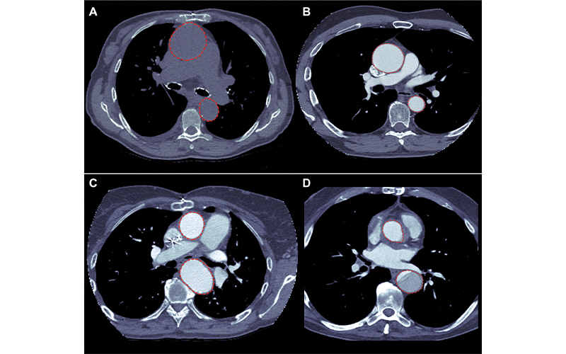
Images of the artificial intelligence model segmentation output (red circles) overlaying the (A) axial CT images of a non–contrast-enhanced CT study with an aneurysm in the ascending aorta, (B) a contrast-enhanced CT study with an aneurysm in the ascending aorta, (C) a contrast-enhanced CT with an aneurysm in the ascending aorta, and (D) a contrast-enhanced CT with dissection in the descending aorta. https://doi.org/10.1148/ryai.210076 ©RSNA 2022
Automatic Detection, Segmentation and Measurement Can Identify Aneurysm
One of those advancements is a fully automatic system for thoracic aorta detection, segmentation and measurement developed and validated by a team of researchers at Massachusetts General Hospital and reported in a Radiology: Artificial Intelligence study.
“The aim of this work is to prevent radiologists from missing an aneurysm in a chest CT performed for non-vascular investigation, especially subtle ones that are not so evident,” Dr. Macruz said, who is one of the study’s lead authors.
The system uses DL to attain the aortic diameters, which many experts report as being a more reliable and reproducible technique than non-AI methodologies.
The AI-model was trained and tested on a dataset consisting of 315 chest and chest plus abdomen CT studies (with and without intravenous contrast enhancement) and CT angiographic studies.
After being linked to other components (a rule-based series filter and a visualization interface), the whole system was validated on another dataset consisting of 1,400 routine CT studies of the chest or chest plus abdomen with and without contrast enhancement.
Extremely Promising Results For Triage and Preventing Underreporting
The system produced good results, particularly when set against a research fellow with two years’ experience in cardiovascular imaging and a radiologist with seven years’ experience with chest CT.
When tested on an independent dataset acquired from a different time period than that used for the main dataset, the system achieved an aneurysm detection accuracy rate of 88% and 81% for the ascending and descending aortas respectively, compared to the research fellow. When compared to the radiologist, this accuracy rate was 90% for the ascending aorta and 82% for the descending aorta.
The mean absolute error between the automatic and the reader-measured diameters was equal to or less than 0.27 cm for both the ascending and descending aortas.
The system is also designed to run silently in the background of PACS, allowing the automatic selection of the axial series with the greatest spatial resolution and, when available, contrast within a complete CT study. Furthermore, the system allows the segmentation of the aorta in the selected series, the measurement of the ascending and descending largest diameters and the visual output of the segmented aortic volume.
“The system produced extremely promising results, not only for triaging TAA-positive studies, but also for preventing underdiagnosis in studies requested for other clinical purposes,” concluded Dr. Macruz.
For More Information
Access the Radiology: Artificial Intelligence study, “Quantification of the Thoracic Aorta and Detection of Aneurysm at CT: Development and Validation of a Fully Automatic Methodology."
Read previous RSNA News stories on AI: