Tiny Lesions Found on US After Thyroid Cancer are Primarily Benign
Research shows that lesions smaller than 6 mm are minimal risk for malignancy

Although it’s common to find small lesions in and around the post-surgical thyroid bed in thyroid cancer patients, most of these lesions are not cancerous, according to a large, retrospective study published in Radiology.
“If tiny lesions are identified and reported on post-thyroidectomy ultrasound, patients and their doctors become understandably anxious and concerned,” said lead author Mary C. Frates, MD, a diagnostic radiologist at Brigham and Women’s Hospital, and professor of radiology at Harvard Medical School, both in Boston.
“Patients worry that the cancer has come back,” Dr. Frates continued. “They want the tiny lesion biopsied and sometimes even removed surgically. But if the lesion is not cancer, that amounts to a lot of unnecessary procedures.”
Dr. Frates, who focuses much of her clinical research on thyroid sonography, said that recent advances in ultrasound (US) equipment are making it possible to find increasingly smaller thyroid bed lesions.
“The goal of this study was to determine if ultrasound can differentiate all the tiny ‘nothings’ discovered in the thyroid bed from the important cancer recurrences,” Dr. Frates said.
Lesion Characteristics and Associations with Malignancy
Dr. Frates and colleagues searched a US reporting database at BWH for all patients imaged between July 2006 and June 2016 with an indication of post-thyroidectomy follow-up.
Researchers analyzed 5,732 US studies in 1,885 patients, 79% of whom were females. Patient age ranged from 3 to 87 years, with a mean age of 48 years.
Recorded data included patient demographic characteristics, date of thyroidectomy, and thyroid cancer type. The vast majority of patients in the study — 82% — had papillary cancer, Dr. Frates said.
The researchers also evaluated the presence, size and US characteristics of thyroid bed lesions, as well as the results of fine-needle aspiration of those lesions (FNA).
Dr. Frates independently reviewed images of all lesions that underwent FNA. She evaluated the shape of thyroid bed lesions, and also looked for echogenicity and the presence of a cystic component, fatty hilum and punctate echogenicities.
Investigators determined that 40% of US examinations showed thyroid bed lesions following thyroidectomy; however, only 2.2% of these lesions proved to be malignant. Furthermore, only 0.2% of lesions in the thyroid bed smaller than 6 mm in maximum diameter were malignant.
“Only the presence of punctate echogenicities had a clear statistical association with malignancy,” Dr. Frates said. “The other characteristics were not helpful in differentiating benign from malignant lesions.”
Patients determined to have positive lymph nodes when they underwent initial thyroidectomy for thyroid cancer had a higher malignancy rate than those who did not have positive lymph nodes at initial surgery.
“Our large study confirms that most patients with thyroid cancer are unlikely to have clinically significant recurrence in the thyroid bed,” Dr. Frates said.
According to the researchers, reporting only lesions 5 mm or larger will simplify US reporting yet allow for appropriate comparison at follow up. Sampling should be considered for lesions 6 mm or larger with concerning features.
“If a lesion does not have punctate echogenicities, it would likely be safe to follow the lesion instead of doing a biopsy,” Dr. Frates said. “But any lesion with punctate echogenicities needs to be taken seriously, and possibly biopsied and removed.”
For More Information
Access the Radiology study, “Role of Sonographic Characteristics of Thyroid Bed Lesions Identified Following Thyroidectomy in the Diagnosis or Exclusion of Recurrent Cancer.”
Read previous RSNA News articles on thyroid cancer:

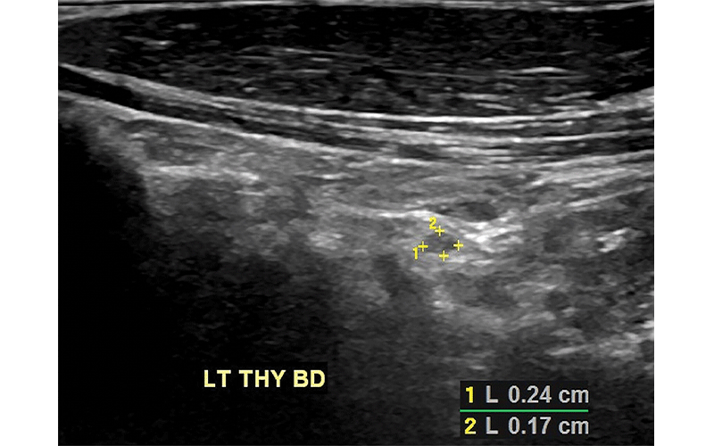
Ultrasound scans of the left thyroid bed (LT THY BD) in a 57-year-old man with history of tall cell variant of papillary thyroid cancer. (a) Sagittal scan shows a 2.4-mm × 1.7-mm solid mass. (b) Transverse scan of the same mass measuring 2.4 mm. This lesion was too small to sample with fine-needle aspiration biopsy but was stable for 5 years and presumed benign.
Frates et al, Radiology 2021© RSNA 2021