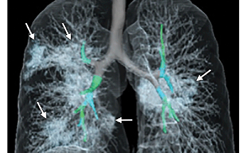
Radiologists Describe Coronavirus Effect on the Lungs
What radiologists should know about the outbreak of new respiratory illness
On Dec. 31, 2019, the World Health Organization (WHO) learned of several cases of a respiratory illness clinically resembling viral pneumonia and manifesting as fever, cough, and shortness of breath. The newly discovered virus emerging from Wuhan City, Hubei Province of China, was temporarily named “novel coronavirus” (2019-nCoV). It is now known officially as COVID-19. This new coronavirus belongs to a family of viruses that include Severe Acute Respiratory Syndrome (SARS) and Middle East Respiratory Syndrome (MERS).
The outbreak is escalating quickly, with hundreds of thousands of confirmed COVID-19 cases reported globally. Early disease recognition is critical not only for prompt treatment, but also for patient isolation and effective public health containment and response.
RSNA has gathered peer-reviewed cases of COVID-19 to provide the global radiology community with a free diagnostic resource to help prevent the spread of this outbreak.
RSNA Resources
- Radiology and Radiology: Cardiothoracic Imaging are publishing original research about coronavirus in a Special Focus section. There is also a Spectrum of Imaging Findings flip-through slide show featuring the images and captions from the available Radiology and Radiology: Cardiothoracic Imaging studies.
- Mar. 10 - Performance of radiologists in differentiating COVID-19 from viral pneumonia on chest CT (Published in Radiology)
- Mar. 6 - FDG PET/CT of COVID-19 (Published in Images in Radiology)
- Feb. 27 - Essentials for Radiologists on COVID-19: An Update - Radiology Scientific Expert Panel (Published in Radiology)
- Feb. 26 - Correlation of Chest CT and RT-PCR Testing in Coronavirus Disease 2019 (COVID-19) in China: A Report of 1,014 Cases (Published in Radiology)
- Feb. 21 - Coronavirus Disease 2019 (COVID-19): A Perspective from China (Published in English and Chinese in Radiology)
- Feb. 20 - CT Chest Findings in Coronavirus-19 (COVID-19): Relationship to Duration of Infection (Published in Radiology)
- Feb. 19 - Sensitivity of Chest CT for COVID-19: Comparison to RT-PCR (Published in Radiology)
- Feb. 13 - Time Course of Lung Changes on Chest CT During Recovery from 2019 Novel Coronavirus (COVID-19) Pneumonia. (Published in Radiology)
- Feb. 13 - Imaging Profile of the COVID-19 Infection: Radiologic Findings and Literature Review. Published in Radiology: Cardiothoracic Imaging)
- Feb. 13 - Chest Imaging Appearance of COVID-19 Infection (Published in Radiology: Cardiothoracic Imaging)
- Feb. 12 - Chest CT for Typical 2019-nCoV Pneumonia: Relationship to Negative RT-PCR Testing. (Published in Radiology)
- Feb. 7 - CT Manifestations of Two Cases of 2019 Novel Coronavirus (2019-nCoV) Pneumonia. (Published in Radiology)
- Feb. 6 - Emerging Coronavirus 2019-nCoV Pneumonia. (Published in Radiology)
- Feb. 4 - CT Imaging Features of Wuhan Coronavirus Infection (2019-nCoV). (Published in Radiology)
· Radiology and Radiology: Cardiothoracic Imaging are publishing editorials and commentaries on COVID-19.
o Feb. 14 – Radiology: Cardiothoracic Imaging Editorial The Many Faces of COVIC-19: Spectrum of Imaging Manifestations, by Fernando Kay, MD and Suhny Abbara, MD. Dr. Abbara is the editor of Radiology: Cardiothoracic Imaging.
o Feb. 4 – Radiology Commentary Chest CT Findings in 2019 Novel Coronavirus (2019-nCoV) Infections from Wuhan, China: Key Points for the Radiologist, by Jeffrey P. Kanne, MD, from the University of Wisconsin School of Medicine and Public Health, Madison.
- Podcasts
- March 2 - Listen to the Radiology podcast with David A. Bluemke, MD, PhD, editor of Radiology, who discusses the latest research published in Radiology.
- Feb. 19 - Listen to the Radiology podcast with David A. Bluemke, MD, PhD, editor of Radiology, who gives an update on COVID-19.
- Feb. 4 - Listen to the Radiology podcast with David A. Bluemke, MD, PhD, editor of Radiology, who discusses Dr. Kanne’s editorial on COVID-19.
- Feb. 12 - Pre and Posttreatment Chest CT Findings: 2019 Novel Coronavirus (2019-nCoV) Pneumonia
- Feb. 12 - Use of Chest CT in Combination with Negative RT-PCR Assay for the 2019 Novel Coronavirus but High Clinical Suspicion
- Feb. 7 - Evolution of CT Manifestations in a Patient Recovered from 2019 Novel Coronavirus (2019-nCoV) Pneumonia in Wuhan, China
- Feb. 4 - 2019 Novel Coronavirus (2019-nCoV) Pneumonia.
- Jan. 31 - CT Imaging of the 2019 Novel Coronavirus (2019-nCoV) Pneumonia.
Images in Radiology: Cardiothoracic Imaging has published:
- Mar. 6 - Severe COVID-19 Pneumonia: Assessing Inflammation Burden with Volume-rendered Chest CT
- Feb. 14 - Severe Acute Respiratory Disease in a Huanan Seafood Market Worker: Images of an Early Casualty
- Feb. 14 - Longitudinal CT Findings in COVID-19 Pneumonia: Case Presenting Organizing Pneumonia Pattern
- Feb. 13 - Spectrum of Chest CT Findings in a Familial Cluster of COVID-19 Infection.
- Feb. 13 - COVID-19 Infection Presenting with CT Halo Sign.

WHO Information
The complete clinical picture with regard to COVID-19 is still not fully clear. Current symptoms reported for patients with COVID-19 have included mild to severe respiratory illness with fever, cough, and difficulty breathing. Coronaviruses can sometimes cause lower-respiratory tract illnesses, such as pneumonia or bronchitis. This is more common in people with cardiopulmonary disease, people with weakened immune systems, infants and older adults. Learn more about the symptoms associated with COVID-19.
WHO has provided information and guidance about the outbreak, including FAQs, situation reports, research updates and technical guidance.