Artificial Intelligence Shows Potential for Triaging Chest X-rays
Chest X-rays account for 40 percent of all diagnostic imaging worldwide.

AI System Developed to Identify Key Findings
For the study, Professor Montana and colleagues used 470,388 adult chest X-rays to develop an AI system that could identify key findings. The images had been stripped of any identifying information to protect patient privacy. The radiologic reports were pre-processed using Natural Language Processing (NLP). For each X-ray, the researchers’ in-house system required a list of labels indicating which specific abnormalities were visible on the image.
“The NLP goes well beyond pattern matching,” Dr. Montana said. “It uses AI techniques to infer the structure of each written sentence; for instance, it identifies the presence of clinical findings and body locations and their relationships. The development of the NLP system for labeling chest X-rays at scale was a critical milestone in our study.”
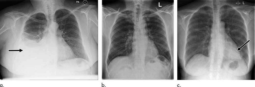
The NLP analyzed the radiologic report to prioritize each image as critical, urgent, non-urgent or normal. An AI system for computer vision was then trained using labeled X-ray images to predict the clinical priority from appearances only. The researchers tested the system’s performance for prioritization in a simulation using an independent set of 15,887 images.
AI System Can Reduce Radiologist’s Workload During Chest X-ray Review
The AI system distinguished abnormal from normal chest X-rays with high accuracy. Simulations showed that critical findings received an expert radiologist opinion in 2.7 days, on average, with the AI approach—significantly sooner than the 11.2-day average for actual practice.

Web Extras
Access the Radiology study, “Automated Triaging of Adult Chest Radiographs with Deep Artificial Neural Networks,” at pubs.radiology.org.