RSNA/AAPM Symposium Addresses Integrated Diagnostics at RSNA 2019
Session will focus on the integration of radiology, pathology and genomics in cancer diagnosis and care


Integrated diagnostics is a familiar topic for radiologists. Radiology training and practice has always been closely linked to pathology and laboratory medicine. While still a nascent field with multiple opportunities for development and exploration, integrated diagnostics can provide a wide variety of potential benefits to patients and give radiologists the opportunity to become more involved in clinical decision support beyond imaging data.
The current and future state of integrated diagnostics will be discussed at the RSNA/American Association of Physicists in Medicine (AAPM) Symposium, “Integrated Diagnostics: Why Does it Matter and How Do We Get There?” at RSNA 2019.
“The development of new and more sophisticated approaches to diagnostic testing, including medical imaging, anatomic pathology and laboratory medicine, along with the growth in targeted cancer therapies, is transforming the landscape of cancer diagnosis and care,” said Paul E. Kinahan, PhD, vice chair for radiology research and head of the imaging research laboratory at the University of Washington, Seattle. “The symposium will help radiologists understand how new methods to combine imaging and other data from multiple diagnostic approaches will ultimately benefit patients and improve outcomes,” said Dr. Kinahan, who will moderate the symposium on Tuesday, Dec. 3.
Using Tools and Data to Aid Diagnosis
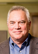
Mitchell Schnall, MD, PhD, will discuss how radiology can be empowered to embrace integrated diagnostics through tools, standards and data science during his symposium presentation, “The Path to Integrated Diagnostics.”
“Although the integrated medical record is often criticized for its impact on operations, it has created an opportunity to improve patient diagnosis by providing access to a broad spectrum of information on each patient,” said Dr. Schnall, the Eugene Pendergrass Professor of Radiology at the Perelman School of Medicine at the University of Pennsylvania, Philadelphia. “As a result, radiologists have access to more non-imaging data than we have ever had before. Integrated diagnostics is slowly becoming the norm of radiology practice, as we integrate laboratory data with our imaging data and clinical findings in our daily work.”
Dr. Schnall acknowledges that current approaches to finding data in medical records are cumbersome and time consuming. While there are few standard approaches to integrating the information, the hope is that new standards will evolve to help radiology to improve diagnostics.
To address these gaps, Dr. Schnall will be leading a new Diagnostic Cockpit Initiative launched by the Academy for Radiology & Biomedical Imaging Research. “This new task force is focused on identifying opportunities for stakeholders and experts to fill the gaps in the development of integrated diagnostics,” Dr. Schnall said.
Cross-Disciplinary Implementation of Integrated Diagnostics
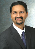
Anant Madabhushi, PhD, will discuss how the development of knowledge in artificial intelligence (AI) and data science has created opportunities to extract and combine information from imaging, pathology, laboratory medicine and genomics during his presentation, “Radio-Patho-Genomics: Computationally Integrated Disease Specific Features Across Scales.”
“There is a need for establishing high fidelity ground truth for disease extent on the radiographic imaging to be able to train AI models,”said Dr. Madabhushi, the F. Alex Nason Professor II of Biomedical Engineering at Case Western Reserve University and a research scientist at the Louis Stokes Cleveland Veterans Administration Medical Center, both in Cleveland. “However granular, ‘ground truth’ definition of disease extent is only available on surgical pathology specimens. AI can help in co-registering the ex-vivo specimens with pre-operative imaging, such as rectal, prostate, lung or breast.”
AI can also be used to create predictors for identifying patients who are likely to have disease recurrence, progression or metastasis, according to Dr. Madabhushi, who has used a combination of AI extracted features from pathology images and CT scans to allow for better recurrence prediction of early stage lung cancer.
The ability to link and associate genomic and molecular information with radiographic patterns on imaging is also a future benefit of integrated diagnostics, Dr. Madabhushi said.
For More Information
The RSNA/AAPM Symposium is part of the Tuesday Morning Plenary at RSNA 2019.