Abnormal Brain Connections Seen in Preschoolers with Autism
Researchers identify altered patterns of global and local brain network topography in young children with ASD

Preschoolers with autism spectrum disorder (ASD), have abnormal connections between certain networks of their brains that can be seen using diffusion tensor imaging (DTI), according to a new study in Radiology. Researchers said the findings may one day help guide treatments for ASD.
ASD refers to a group of developmental disorders characterized by communication difficulties, repetitive behaviors and limited interests or activities. ASD can usually be diagnosed within the first few years of life. Early diagnosis and intervention are important because younger patients typically benefit most from treatments to improve their symptoms and ability to function.
For the study, researchers compared DTI results between 21 preschool boys and girls with ASD (mean age of 4-and-a-half years old) with those of 21 similarly aged children with typical development. Applying graph theory to the DTI results, the researchers measured the relationships among highly connected areas of the brain.
“While developments in brain imaging have enabled the discovery of abnormal brain connectivity in younger children with ASD, the phenomenon has not yet been fully investigated at the brain network level,” said study co-author Lin Ma, MD, Chinese PLA General Hospital, Beijing.
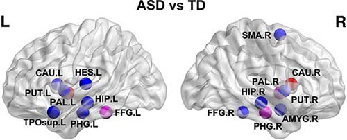
Compared with the typically developed group, children with ASD demonstrated significant differences in components of the basal ganglia and paralimbic-limbic networks, systems that play a crucial role in behavior.
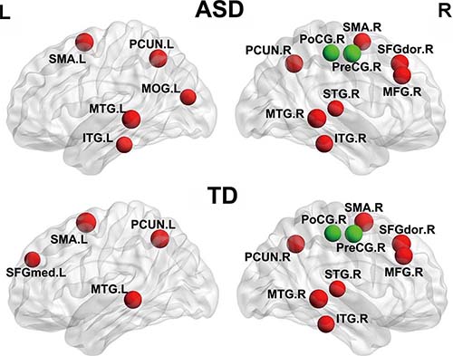
Global topologic patterns of white matter networks in children ASD were significantly altered, as indicated by a shortened characteristic path length and increased global efficiency and clustering coefficient.
Nodes with significant topologic alternations were mainly distributed in the basal ganglia network, including the bilateral caudate, putamen and pallidum and in the paralimbic-limbic network, including the bilateral hippocampus, parahippocampal gyrus, left temporal pole of the superior temporal gyrus and right amygdala.
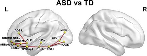
“The observed increase in connections between the paralimbic-limbic system and the occipital network demonstrated the important role of the paralimbic-limbic system in childhood ASD,” Dr. Ma said. “Disconnections between these networks may be associated with an aberrant observation and imitation capacity in childhood ASD.”
The researchers also observed areas of decreased nodal efficiency in the supplementary motor area in ASD, which may be associated with motor dysfunction.
In addition, significantly altered white matter connectivities were located in the left hemisphere, which might provide support for the left-hemisphere hypothesis for autism, which provides explanations for many symptoms of autism, such as language deficits and delayed language development.
“There were correlations between the white matter networks topologic alterations and the clinical scales of the Childhood Autism Rating Scale, suggesting delayed or stunted maturation processes in the children with ASD,” Dr. Ma said.
The identification of altered structural connectivity in these networks may point toward potential imaging biomarkers for preschool children with ASD.
“The imaging finding of those ‘targets’ may be a clue for future diagnosis and even for therapeutic intervention in preschool children with ASD,” Dr. Ma said.


Web Extras
- Access the study, “Alterations of White Matter Connectivity in Preschool Children with Autism Spectrum Disorder,” at https://pubs.rsna.org/doi/full/10.1148/radiol.2018170059