Weight Loss Can Slow Down Knee Joint Degeneration

Overweight and obese patients who lost a substantial amount of weight over a 48-month period showed significantly lower degeneration of their knee cartilage, according to new Radiology research.
Researchers at the University of California, San Francisco, looked at the degeneration of menisci, articular cartilage and bone marrow in the knee joint, according to the study’s lead author, Alexandra Gersing, MD, Department of Radiology and Biomedical Imaging.

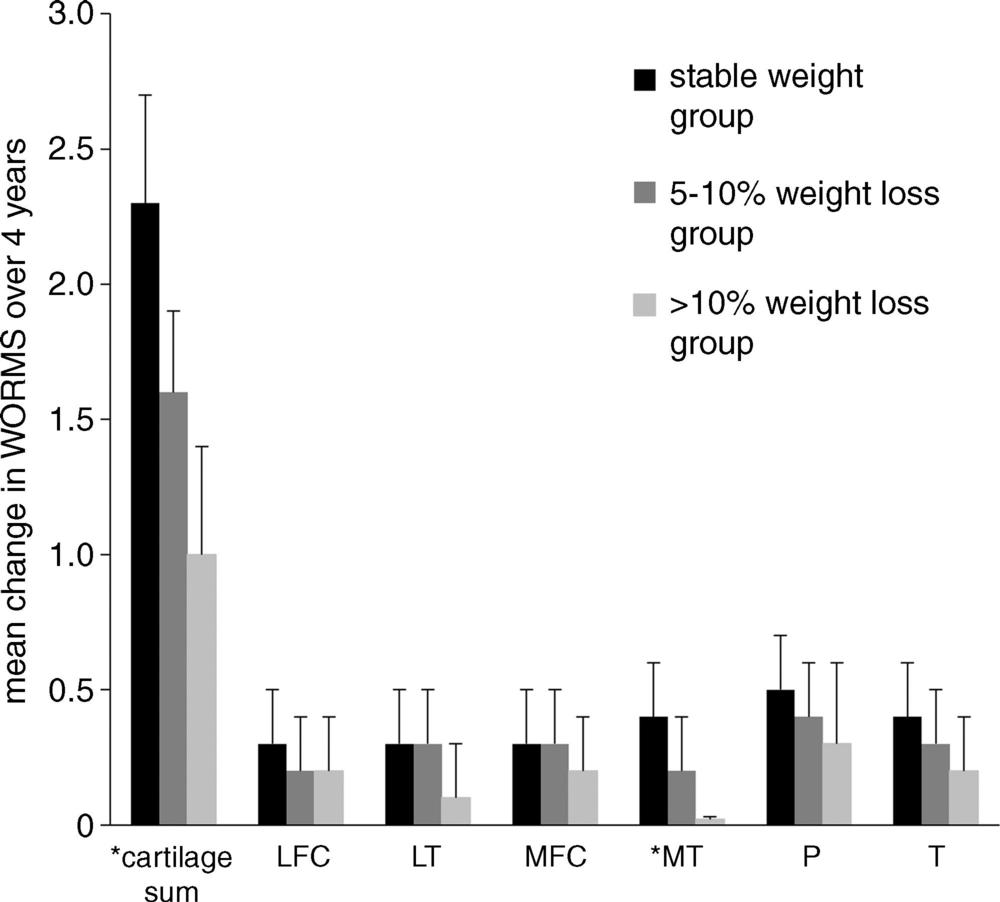
Dr. Gersing and colleagues investigated the association between weight loss and the progression of cartilage changes on MRI over a 48-month period in 640 overweight and obese patients (minimum body mass index [BMI] 25 kg/m2) who had risk factors for osteoarthritis or MRI evidence of mild to moderate osteoarthritis using data that was collected from the Osteoarthritis Initiative. Patients were categorized into three groups: those who lost more than 10 percent of their body weight, those who lost five to 10 percent of their body weight and a control group whose weight remained stable.
The results showed that patients with five percent weight loss had lower rates of cartilage degeneration when compared with stable weight participants. In those with 10 percent weight loss, cartilage degeneration slowed even more.

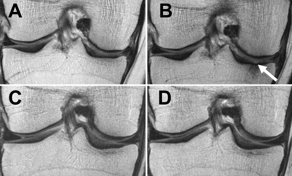
In addition to slowed articular cartilage degeneration, researchers also saw changes in the menisci after weight loss.
“The most exciting finding of our research was that not only did we see slower degeneration in the articular cartilage, we saw that the menisci degenerated a lot slower in overweight and obese individuals who lost more than five percent of their body weight, and that the effects were strongest in overweight individuals and in individuals with substantial weight loss,” Dr. Gersing said.
Web Extras
- Access the Radiology study “Is Weight Loss Associated with Less Progression of Changes in Knee Articular Cartilage among Obese and Overweight Patients as Assessed with MR Imaging over 48 Months? Data from the Osteoarthritis Initiative” at pubs.rsna.org