Journal Highlights
The following are highlights from the current issues of RSNA’s two peer-reviewed journals


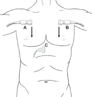
Focused Assessment with Sonography in Trauma (FAST) in 2017
The advent of focused assessment with sonography in trauma (FAST) three decades ago enabled clinicians to rapidly screen for injury at the bedside of patients, especially those patients too hemodynamically unstable for transport to the CT suite.
In the April issue of Radiology (RSNA.org/Radiology), John R. Richards, MD, and John P. McGahan, MD, from the University of California-Davis Medical Center, discuss the evolution of the FAST examination to its current state in 2017 and evaluate its evolving role in the acute management of the trauma patient.
The authors also report on the utility of FAST in special patient populations, such as pediatric and pregnant trauma patients, and the potential for future research, applications and portions of this examination that may be applicable to radiology-based practice.
“The most effective use of focused assessment with sonography in trauma has been rapid triage of hemodynamically unstable trauma patients to definitive intervention, leading to reduced time to appropriate intervention, shortened hospital stays and lower costs,” the authors write.
This article meets the criteria for AMA PRA Category 1 Credit™. SA-CME is available online only.
Multidetector CT of Surgically Proven Blunt Bowel and Mesenteric Injury
Blunt bowel and mesenteric injury is relatively uncommon in the setting of blunt abdominal trauma. However, a timely diagnosis is paramount to the proper triage and management of trauma patients.
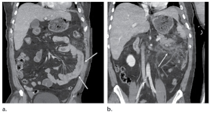
In the March-April issue of RadioGraphics (RSNA.org/RadioGraphics), David D. B. Bates, MD, of Boston University Medical Center, and colleagues identify and describe three mechanisms of blunt bowel and mesenteric injury; discuss the sensitivity and specificity of various CT findings of blunt bowel and mesenteric injury; and discuss ways to recognize potential pitfalls in the evaluation of patients who are suspected of having blunt bowel and mesenteric injury.
Despite the relatively low rate of blunt bowel and mesenteric injury in patients with abdominal and pelvic trauma, delays in diagnosis are associated with increased rates of sepsis, a prolonged course in the intensive care unit and increased mortality.
“Once radiologists are familiar with the spectrum of findings of blunt bowel and mesenteric injury, they will be able to make timely diagnoses that will lead to improved patient outcomes,” the authors write.
This article has an Invited Commentaryby Michael N. Patlas, MD, Department of Radiology, Hamilton General Hospital, Hamilton, Ontario, Canada.”
This article meets the criteria for AMA PRA Category 1 Credit™. SA-CME is available online only.
Radiology Podcasts
 Listen to Radiology Editor Herbert Y. Kressel, MD, deputy editors and authors discuss the following articles in the February issue of Radiology at RSNA.org/Radiology-Podcasts.
- “Impact of Medicare Shared Savings Program Accountable Care Organizations at Screening Mammography: A Retrospective Cohort Study,” Anand K. Narayan, MD, and colleague.
- “Lesion Topography and Microscopic White Matter Tract Damage Contribute to Cognitive Impairment in Symptomatic Carotid Artery Disease,” Dewen Meng, MSc, and colleagues.
RadioGraphics Podcasts
 Listen to RadioGraphics Editor Jeffrey S. Klein, MD, and authors discuss the following article in the March-April 2017 issue of RadioGraphics at pubs.RSNA.org/Page/RadioGraphics/Views.
- “Multidetector CT of Surgically Proven Blunt Bowel and Mesenteric Injury,” by David D.B. Bates, MD, and colleagues.

