Disruption in Brain’s Communication Networks Key to Vascular Cognitive Disorder
Subcortical white matter ischemic lesion locations and severity of ultrastructural tract damage contribute to cognitive impairment in symptomatic Caratoid Artery Disease (CAD), which suggests that subcortical disconnection within large-scale cognitive neural networks is a key mechanism of vascular cognitive disorder, according to new Radiology research.
In a multimodal MR imaging study of 108 patients with symptomatic CAD but no dementia, Dewen Meng, MSc, of the University of Nottingham, England, and colleagues conducted comparisons and interrelations between global cognitive and fluency performance, lesion topography and ultrastructural damage, assessed with voxel-based statistics. Associations between cognition, medial temporal lobe atrophy lesion volumes, and global white matter ultrastructural damage indexed as increased mean diffusivity were tested with regression analysis by controlling for age.
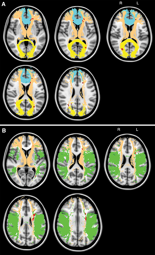
In patients with CAD, researchers determined that cerebrovascular lesion location in the thalamic radiation and interhemispheric fiber tracts contributes to global cognitive deficits and lesions in the tha-lamic radiation and long association fibers affect fluency performance. Severity of subcortical tissue damage — preferentially in major white matter tracts — contributes to global cognitive impairment with skeleton mean diffusivity as the best-performing imaging marker for the prediction of probable vascular cognitive disorder, researchers determined.
“The findings provide insights into how cerebrovascular disease contributes to cognitive impairment and/or dementia and highlight the need to combat progressive subcortical brain tissue damage; average white matter skeleton mean diffusivity shows potential as a simple diagnostic marker of subcortical disconnection underlying vascular cognitive disorder,” the authors write.
