Imaging Predicts Long-Term Effects in Veterans with Brain Injury

Differences in white matter microstructure may partially account for the variance in functional outcomes among veterans who sustained combat-related mild traumatic brain injury (TBI), according to new research.
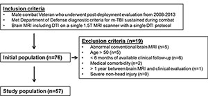
Jeffrey B. Ware, M.D., of the Philadelphia VA Medical Center, and colleagues studied 57 male combat veterans with mild TBI who had undergone brain MRI between Jan. 1, 2008, and Sept. 30, 2013.
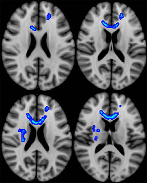
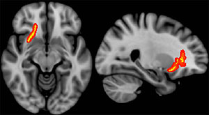
Overall, veterans had a mean of 46 health care visits per year during the follow-up period. Cumulative health care visits over time were inversely correlated with diffusion anisotropy of the splenium of the corpus callosum and adjacent parietal white matter. Clinical measures obtained during initial postdeployment evaluation were not predictive of later functional status.
“Our results suggest that diffusion measurements hold the potential to confer important prognostic information in the clinical evaluation of combat-related mild TBI. These findings should encourage continued research efforts to further refine and investigate the clinical utility of diffusion measurements in this setting,” the authors write.


Web Extras
- Access the study, "Combat-related Mild Traumatic Brain Injury: Association between Baseline Diffusion-Tensor Imaging Findings and Long-term Outcomes," at pubs.rsna.org/doi/10.1148/radiol.2016151013