Radiology Imaging Education Focus of International Panel
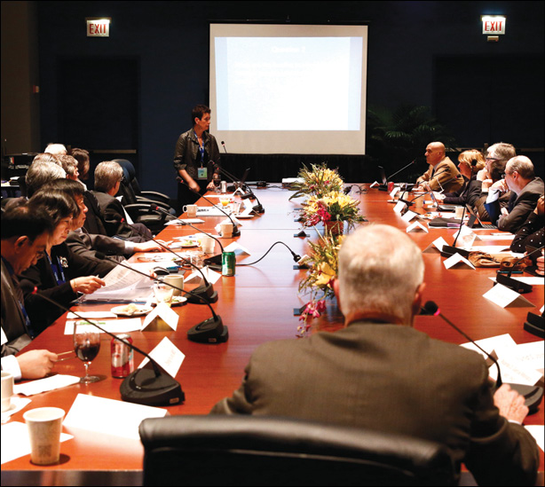
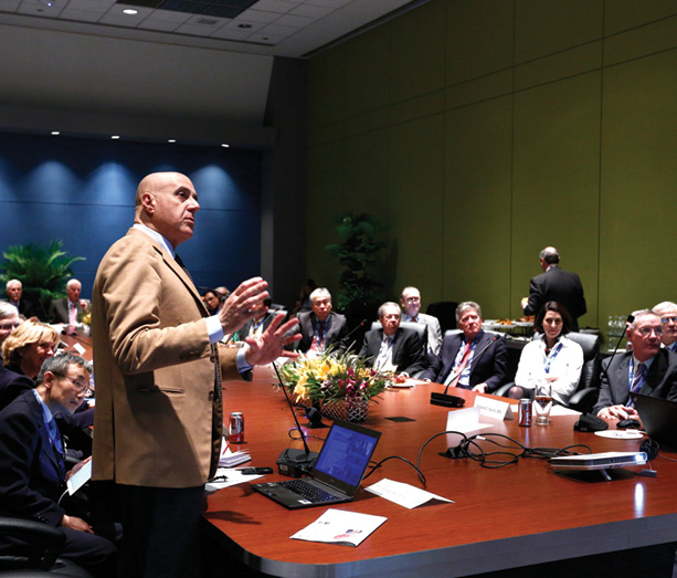
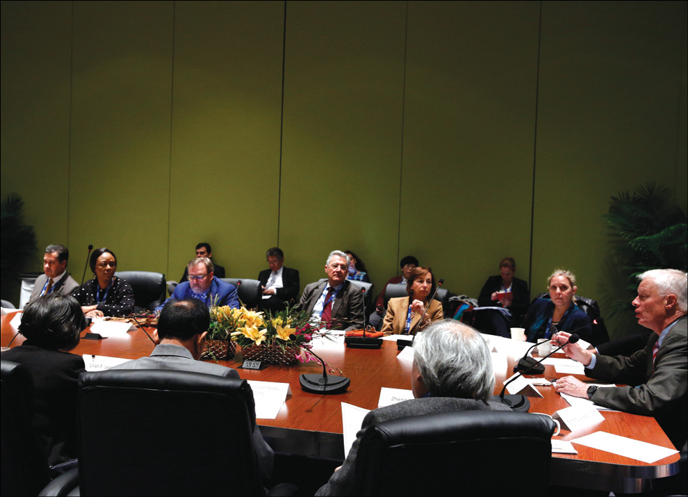
Experts from across the globe discussed education the generation of radiologists at the International Trends Session held at RSNA 2015
While each brought a different set of experiences to the table, a panel of experts from around the globe also found a lot of common ground in their approach to educating the next generation of radiologists during the International Trends Session held at RSNA 2015.
Radiology education should be interesting and interactive, incorporate technology and e-learning, be simple when possible, and above all, be patient-centered, according to presenters gathered to discuss “International Medical Student Imaging Education.”
Each year the International Trends meeting is held to bring radiology organizations together to share ideas and best practices on a topic of global importance to the profession.
In discussing the basics of teaching medical students about radiologic equipment and radiology protection in the U.K., Christiane Nyhsen, M.D., consultant radiologist at City Hospitals Sunderland NHS Foundation Trust and senior lecturer at Newcastle University, stressed the importance of keeping students’ attention.
“Radiology protection needs to be presented to students in an interesting way,” Dr. Nyhsen said. “Share the challenges of what low-dose radiation risks may or may not be. Tell them about studies in geographic areas of high background radiation. Mention radiation spas, emerging evidence of how the human body responds to low levels of radiation. It is quite fascinating in my opinion.”
Radiological equipment is best taught alongside real cases, Dr. Nyhsen said. Interactive discussions help keep students interested. For example, learning points on MRI safety can be nicely integrated between case discussions, she said.
“Teach medical students well and future doctors will have a much better understanding of how to make the best use of imaging modalities available,” she added.
And make sure to put patients first.
“Put the patient in the center of teaching,” Dr. Nyhsen said. “Imaging should help the patient—the patient should be considered, advised, have tests appropriately explained and their fears reduced.”
Medical Imaging Should be Taught on Day One
Petra Lewis, M.B.B.S., discussed the introduction of medical imaging into anatomy and more specifically, offered her opinion on when it should be introduced, why it should be incorporated, who should teach it, and how it should be taught.
“The ‘when’ is easy,” said Dr. Lewis, of the Geisel School of Medicine at Dartmouth, in Hanover, New Hampshire. “It should start on Day One of the anatomy course and preferably Day One of medical school.”
Medical imaging should be incorporated early into preclinical curriculum since all medical students will use it either directly or indirectly their entire careers. Radiologists should be teaching anatomy as well as medical imaging, Dr. Lewis said.
“We know the anatomy, the imaging and the clinical impact of these findings,” Dr. Lewis said. “Residents and fellows are fabulous at teaching anatomy and usually greatly enjoy the experience. Use the resources you have available to teach.”
All modalities can be used to teach anatomy depending on the organ system, Dr. Lewis said. Online modules that already exist or are created within the institution can help, especially for pre-learning materials. Short videos are easy to make and are effective, she said.
Keep it Simple, Use E-learning
Simplification is the key to introducing the basics of medical imaging technology, dose and safety, said Francesco Sardanelli, M.D., a professor of radiology at the University of Milan, Italy.
“We should try to reduce complexity to simplicity,” Dr. Sardanelli said. “We need to present the meaning of words we use, to explain to students the risk of the MR environment and translate the numbers to something easy to understand. One example is better than 20 lines of text.”
In Japan, lecture-theatre style education is still dominant in radiology, said Kunihiko Fukuda, M.D., a professor of radiology at the Jikei University School of Medicine in Tokyo. However, “It is desirable to create more e-learning materials and use them effectively in the flipped education style,” Dr. Fukuda said. “If the lecture style will be continued, radiology should be integrated with clinical medicine and pathology.”
Medical education is six years in Japan, Dr. Fukuda said. Radiation physics and biology start at early stages in lecture style, he said, while imaging diagnosis and radiation oncology start usually from year four. “They are usually started with the lecture style and then progress to a tutorial and/or elective course,” Dr. Fukuda said.


