MRI Improves Diagnosis of Microbleeding after Brain Injury in Military Personnel
Performing MR imaging in patients with traumatic brain injury (TBI) as early as possible would facilitate better evaluation of cerebral microhemorrhages (CMHs), according to new research published online in the journal, Radiology.
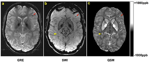
Wei Liu, D.Sc., of Walter Reed National Military Medical Center, and colleagues studied 603 military personnel patients who underwent two-dimensional conventional gradient-recalled-echo MR imaging and three-dimensional flow-compensated multiecho gradient-recalled-echo (GRE) MR imaging (processed to generate susceptibility-weighted images [SWI] and quantitative susceptibility maps [QSM]), and a subset of patients who underwent follow-up imaging. Microhemorrhages were identified by two radiologists independently. Comparisons of microhemorrhage number, size, and magnetic susceptibility derived from quantitative susceptibility maps between baseline and follow-up imaging examinations were performed by using the paired t test.
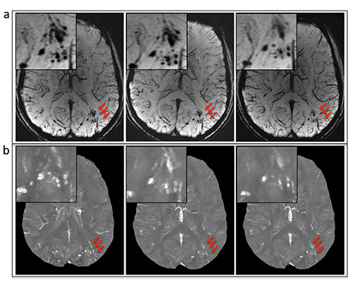
Among the 603 patients, cerebral microhemorrhages were identified in 43 patients, with six excluded for further analysis owing to artifacts. Seventy-seven percent (451 of 585) of the microhemorrhages on susceptibility-weighted images had a more conspicuous appearance than on gradient-recalled-echo images. Thirteen of the 37 patients underwent follow-up imaging examinations. In these patients, a smaller number of microhemorrhages were identified at follow-up imaging compared with baseline on quantitative susceptibility maps (mean ± standard deviation, 9.8 microhemorrhages ± 12.8 vs. 13.7 microhemorrhages ± 16.6; P = .019). Quantitative susceptibility mapping–derived quantitative measures of microhemorrhages also decreased over time: -0.85 mm3 per day ± 1.59 for total volume (P = .039) and -0.10 parts per billion per day ± 0.14 for mean magnetic susceptibility (P = .016).
“In the current study we found that the number of microhemorrhages and QSM-derived measurements of microhemorrhages all decreased over time, suggesting that hemosiderin products undergo continued, subtle evolution in the chronic stage of TBI. Furthermore, by correlating microhemorrhages with regional brain volumes, abnormalities such as fiber discontinuities or hyperintensities on T2-weighted fluid attenuation inversion-recovery images will facilitate the investigation of this disease,” the authors write.


Web Extras
- Access the study, "Imaging Cerebral Microhemorrhages in Military Service Members with Chronic Traumatic Brain Injury," at pubs.rsna.org/doi/full/10.1148/radiol.2015142554