Future of 3-D Printing is Bright, but Cost Remains an Obstacle
Along with a significant impact on face transplantation, 3D printing is providing remarkable results in planning complex, life-changing surgeries


With advanced imaging as the cornerstone, the future of 3-D printing is bursting with possibilities for all of healthcare and holds the potential to completely reshape medicine. But barriers—primarily training opportunities and cost—prevent the technology from becoming widely implemented in everyday practice anytime soon.
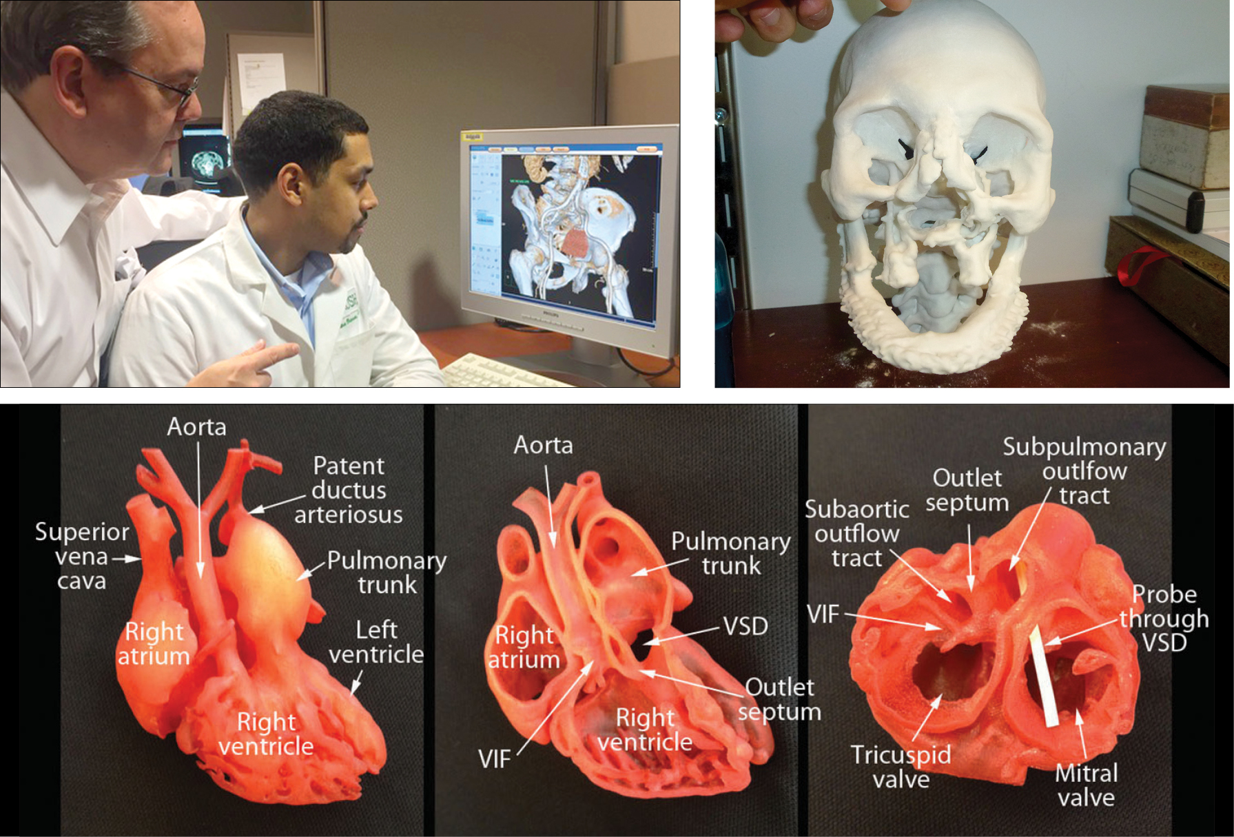
Already 3-d printing is providing remarkable results in planning complex interventions in the heart, the spine and for a growing list of other procedures. At RSNA 2014, a research team led by Frank J. Rybicki, M.D., Ph.D., former director of the Applied Imaging Science Lab at Brigham and Women’s Hospital (BWH) in Boston, and incoming chair and professor at the University of Ottawa, Canada, demonstrated how they utilized CT and 3-D printing to recreate life-size models of patients’ skulls and soft tissues to assist in face transplantation surgery.
In 2011, Dr. Rybicki, working with the BWH surgical team led by Bohdan Pomahac, M.D., performed the first successful full-face transplantation in the U.S. and the team has since completed four more full-face procedures in addition to other partical face transplants. Dr. Rybicki explained that 3-D visualization was the precursor to 3-D printing and took place in 3-D labs. The underlying idea was that DICOM (Digital Imaging and Communications in Medicine) data sets contained more useful information than was being extracted via axial images alone, or even the rendering of a 3-D volume on a 2-D monitor (the essence of 3-D visualization). The availability of 3-D printing was the next logical step in technology and application to patient care, Dr. Rybicki said.
“There are strong parallels between 3-D printing today and early 3-D labs,” Dr. Rybicki said. “Even more information can be obtained in DICOM images via a printed model in your hands, and this has borne out in many fields.
“3-D printing impacts far more routine surgeries than face transplantation, and it is here to stay,” Dr. Rybicki added.
3D Printing Aids Heart, Spine Surgeons
Advancements in other areas support Dr. Rybicki’s statement. The benefits of using 3-D printing to create a 3-D model of the heart were a major point of emphasis at the EuroEcho-Imaging 2014 annual meeting of the European Association of Cardiovascular Imaging (EACVI) last summer.
Specifically, in fall 2014, a 3-D heart model helped surgeons at Morgan Stanley Children’s Hospital in New York repair a congenital heart defect in a 2-week-old baby. “With the advent of 3-D imaging, now we can clearly evaluate the structure of the heart in different planes,” said Patrizio Lancellotti, M.D., Ph.D., EACVI president, in a written statement. “With this novel technology we will gain insights into the interactions between the valves and the ventricles, the valves and the aorta, and the valves and the left atrium.”
3-D printing itself depends on the advanced imaging modalities and protocols to generate source DICOM images amenable for printing. “For certain applications such as cardiac congenital anomalies, advanced MR protocols have proven invaluable to generate 3-D printed models," Dr. Rybicki said. "For other applications, particularly when bony structures are printed, thin section CT images will suffice.”
In another breakthrough procedure in 2014, 3-D printing was used in complex spinal cord surgery. Doctors from the Peking University Third Hospital in Beijing successfully replaced a section of cancerous vertebra in a 12-year-old boy’s neck with a piece created on a 3-D printer.
3-D printing was also successfully used in at least five other life-changing surgeries during 2014, including replacing an upper jaw, forming a new skull, spinal-fusion surgery, and heel and hip implants.
Training, Cost, Barriers to Widespread Use
Despite these successes, the high cost of 3-D printing hampers its implementation into everyday practice. According to an article in Crain’s Chicago Business, high-resolution printers cost anywhere from $40,000 up to $1 million.
Other factors also run up the costs. For instance, implementing a 3-D printing laboratory is expensive, not only because the hardware and software are costly, but also because the work is very time- and labor-intensive. Funding sources, including federal resources, have become increasingly scarce, which make the funding environment extremely competitive.
In terms of software and hardware, expenses to implement a 3-D printing laboratory, funding has become increasingly difficult as federal resources have decreased, making the process extremely competitive. “Applying for funding used to be a fairly quick process, but now it’s becoming ever more prolonged,” said John F. Hibbeln, M.D., who operates the 3-D printing lab at Rush University Medical Center in Chicago. “In the era of increasing restraint on medical costs, how does this fit in? The real issue is money and who’s going to pay for it.”
Dr. Hibbeln said his institution uses an independent contractor for their 3-D printing rather than buying the equipment. But he believes costs will begin decreasing soon as more companies become involved.
“The price of printing will come down. The number of printers out there is increasing. The utility is increasing. The software is improving. And the prices are dropping with all computers, so I anticipate the overall price will drop,” Dr. Hibbeln said.
Because he works at a teaching hospital, Dr. Hibbeln sees the greatest potential for 3-D printing in areas of education, training and research. He added one other oft-overlooked potential benefit:
“We teach medical students, residents, fellows and junior physicians, but we are also here to teach patients,” he said. “This is another part of the teaching mission that is underestimated. How do patients understand what is going on?"
A 3-D printed model can be extremely effective for physicians seeking informed consent for patients and their families. “When the patient actually holds his actual abnormal anatomy, or the parents hold that anatomy of their child in their hands, they can truly understand the problem and what the physician intends to do to correct the abnormality,” Dr. Rybicki said.
“3-D printing has tremendous potential. Like so much else in the world, the only constraints are money and inventiveness,” Dr. Hibbeln added.
Web extras - Videos
Watch videos of Dr. Rybicki’s RSNA 2014 research on 3-D printing including footage of the animated face transplant process and an RSNA 2014 interview with Dr. Rybicki explaining the 3-D printing process, below.
Footage showing animation of face transplant process on a male patient.
Dr. Frank Rybicki explains why CT is critical to making the 3-D model.
