Ultrasound Aids Carpal Tunnel, Plantar Fasciitis
Radiology research shows the effectiveness of new ultrasound applications in diagnosing carpal tunnel syndrome and chronic plantar fasciitis


Applications for ultrasound continue to grow with one recent study demonstrating the modality’s effectiveness in diagnosing carpal tunnel syndrome (CTS) and another showing its potential in treating chronic plantar fasciitis.
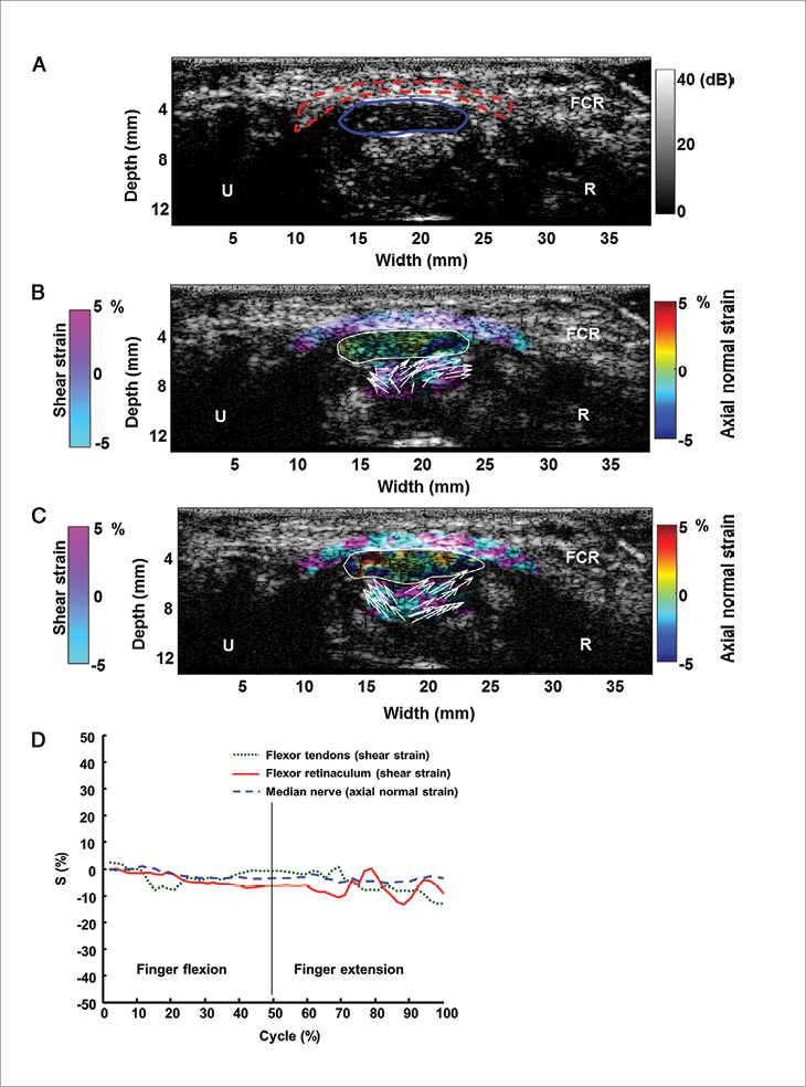
In a study published in the April 2015 issue of Radiology, Taiwanese researchers who developed a new 2D ultrasound strain imaging technique demonstrated its effectiveness in diagnosing CTS, and at an earlier stage than other imaging techniques. The study found that ultrasound strain imaging can quantify and map tissue kinematics effectively enough to detect CTS.
Researchers who studied the wrists of 26 patients (10 healthy and 16 diagnosed with CTS) in real time determined that the standard deviation of the cumulative strain (SDCS) for the shear strain of the flexor retinaculum was significantly lower in patients with CTS than in healthy volunteers. Researchers also determined that the axial strain of the median nerve was higher in healthy volunteers than in patients with CTS.
It was important that the subjects followed a prescribed finger-motion described in the study to properly measure the strain. “Subjects have to practice and follow the physician’s instructions to perform one cycle of standardized finger motion from the neutral posture to maximum flexion and then to full extension during a span of 2-3 seconds,” said study author Chih-Kuang Yeh, Ph.D., a professor and biomedical researcher at National Tsing Hua University in Taiwan.
Combining ultrasound strain imaging with the prescribed finger motion enabled researchers to quantify the SDCS in the problem region. By adding the temporal variation of the tissue deformation within one cycle of active finger motion, they were able to quantify the cyclic carpal tunnel tissue mobility.
Although commercial software of strain imaging is provided by most ultrasound imaging companies, “our proposed technique is easier to implement on available clinical ultrasound platforms. We just apply a color map that corresponds to strain information) and overlap it with a B-mode image to show the ultrasound strain imaging,” Dr. Yeh said.
Ultrasound imaging is not only less expensive and less invasive than MRI, “there are some cases of subclinical CTS or early-stage CTS where electrodiagnostic testing cannot detect median nerve dysfunction,” Dr. Yeh said.
Dr. Yeh added that further research was necessary to confirm their preliminary results due to the small sample size of their study.
Ultrasound Aids Chronic Plantar Fasciitis
In another study published online in February 2015 in the Journal of Vascular and Interventional Radiology, radiologists in Nebraska determined that ultrasonic therapy brought relief to 90 percent of patients with chronic plantar fasciitis. Interventional radiologists at Advanced Medical Imaging, Lincoln, treated 65 patients starting in August 2013 after medication, activity modification, physical therapy and arch supports failed to help patients.
“We have treated well over 100 patients with chronic plantar fasciitis and we are seeing greater than 90 percent improvement,” said Rahul S. Razdan, M.D., the study’s lead author and an interventional radiologist at Advanced Medical Imaging.
Patients in the study were chronic sufferers who had tried other treatments. “Our patients were at their wits end and were planning to resort to surgery,” Dr. Razdan said.
Ultrasound is used in every step of the therapy and is especially crucial for targeting the unhealthy tissue, he said. “The most integral part of the procedure is to know what you’re looking at and seeing where the bad collagen is in real time,” Dr. Razdan said.
According to the study, a percutaneous ultrasonic fasciotomy was performed with ultrasound guidance from a TX1 device. When activated, the device’s hollow 18g needle tip emulsifies tissue using high-frequency, low-amplitude motions, which is then extracted via a pump.
The clinic treats plantar fasciitis as an overuse injury that leaves behind scar tissue that displaces healthy tissue. “Our job is to remove the diseased collagen and to give healthy collagen a chance to deposit,” Dr. Razdan said.
Patients were evaluated based on the Foot and Ankle Disability Index (FADI) pre-procedure and post-procedure at two weeks, six weeks and six months. Dr. Razdan said some of the earliest treated patients are showing no signs of reversal after almost two years.
The ultrasound therapy still must be combined with physical therapy to correct the habits that caused the overuse injury in the first place, Dr. Razdan said. But the treatment can get more people back to the recreation they love or the jobs they need to make a living.
Applications for ultrasound therapy have been expanding to injuries in the elbow, patella and Achilles tendon, he added.
Although not included in the study, Dr. Razdan said data showed a correlation between the duration of the ultrasonic therapy and higher FADI scores, which he attributes to the removal of greater amounts of bad collagen, allowing more blood cells to flow into the area and stimulate healing.
 Radiology Select Focuses on Imaging of Joints
Imaging and joints is the focus of Volume 6 of Radiology Select—a continuing series of articles that highlight developments in imaging science, techniques and clinical practice. Articles in Volume 6 were personally selected by guest editor Thomas M. Link, M.D., Ph.D., a radiology professor in the departments of radiology and biomedical imaging at the University of Muenster, Germany, and cover:
- Anatomy, technique and scanning pitfalls of shoulder ultrasounds
- Bilateral MRI of the hand and wrist from early inflammatory arthritis to rheumatoid arthritis
- Evaluating the articular cartilage of knee joints by adding T2 mapping sequences to routine MR imaging protocol
- MRI for diagnosis and reconstruction of the knee, including clinical importance, possible infection and follow-up
- New imaging developments for hip, pelvis, ankle and foot issues to help identify athletic pubalgia, ligament damage and bursae development
The Online Educational Edition includes 22 tests with an opportunity to earn 22 SA-CME credits. This enduring material can be applied towards the ABR self-assessment requirement.
Radiology Select is available to members for a discounted price at RSNA.org/RadiologySelect.

Web Extras
- Access the study “Carpal Tunnel Syndrome: US Strain Imaging for Diagnosis,” at RSNA.org/Radiology
- Access the study “Percutaneous ultrasonic fasciotomy: a novel approach to treat chronic plantar fasciitis,” at jvir.org