Journal highlights
The following are highlights from the current issues of RSNA’s peer-reviewed journals.
AI vs. Radiologists: Breast Cancer Detection After Mastectomy
Patients with a personal history of breast cancer (PHBC) are at risk for subsequent breast cancer. Annual surveillance mammography is recommended for these patients, but compared with routine mammography in patients without a PHBC, it has lower sensitivity and higher interval cancer rates.
More effective tools are needed to enhance screening and surveillance mammography and improve patient outcomes. AI is a promising approach, but limited data exist regarding the accuracy of AI algorithms in patients with a PHBC treated with unilateral mastectomy.
In a new study published in Radiology, researchers led by Sun Min Ha, MD, PhD, from Seoul National University Hospital, Korea, compared the performance of standalone AI with that of radiologists in detecting contralateral breast cancer in patients after unilateral mastectomy. Standalone AI demonstrated higher sensitivity and cancer detection rates than radiologists without AI assistance.
“However, the relatively high proportion of cancers missed by both AI and radiologists underscores the need for further studies to support the translation of this promising technology into routine clinical practice,” the authors caution.
Read the full article, “Breast Cancer Detection with Standalone AI versus Radiologist Interpretation of Unilateral Surveillance Mammography after Mastectomy.”
Follow the Radiology editor on X @RadiologyEditor.

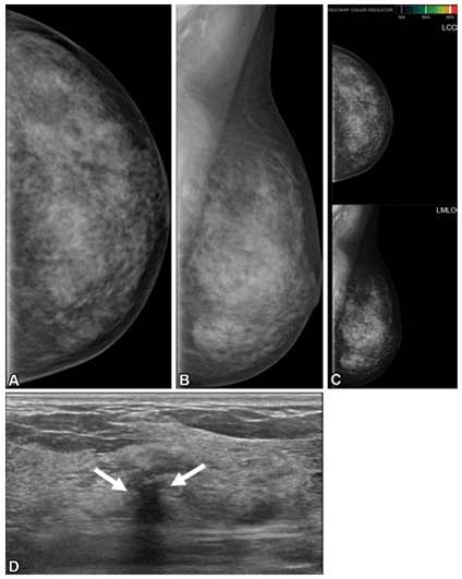
Images in a 51-year-old patient with contralateral second breast cancer 2.8 years after right mastectomy. (A) Left craniocaudal and (B) mediolateral oblique mammograms assessed as negative. (C) When AI was applied, no abnormalities were noted. (D) Supplemental axial US scan, which was obtained on the same day as the mammogram, shows an irregular hypoechoic mass (arrows) in the left upper outer breast. Pathology results at biopsy revealed invasive ductal carcinoma (pT1N0, 1.0 cm, estrogen receptor negative, progesterone receptor negative, human epidermal growth factor receptor type 2 negative, histologic grade II). LCC = left craniocaudal, LMLO = left mediolateral oblique.
https://doi.org/10.1148/radiol.242955 ©RSNA 2025
Understanding Hip Microinstability
Hip instability has been traditionally associated with trauma, hip arthroplasty or developmental dysplasia. Recently, the term microinstability has emerged to describe more subtle forms of hip instability. Unlike classic instability, microinstability does not involve dislocation but causes pain and discomfort, particularly when descending stairs.
Microinstability is commonly seen in young or middle-aged patients, especially women and athletes. Radiologists play a key role in early detection to help prevent chondrolabral injury and premature osteoarthrosis.
A recent RadioGraphics article offers a comprehensive guide to the evolving concepts and definitions of hip instability. Júlio B. Guimarães, MD, PhD, from Fleury Medicina e Saúde in São Paulo, and colleagues describe bone and joint anatomy as well as biomechanics of the hip. They also emphasize the role of imaging—like leg traction MRI and dynamic US—in detecting elusive signs that are often missed on physical examination or screening radiographs.
“The radiologist should be able to apply this knowledge in their multidisciplinary hip practice to aid timely diagnosis and management of such disorders,” the authors conclude.
Read the full article, “Hip Microinstability: Novel Concepts and Comprehensive Imaging Evaluation.” This article is also available for CME on EdCentral. Follow the RadioGraphics editor on X @RadG_Editor.

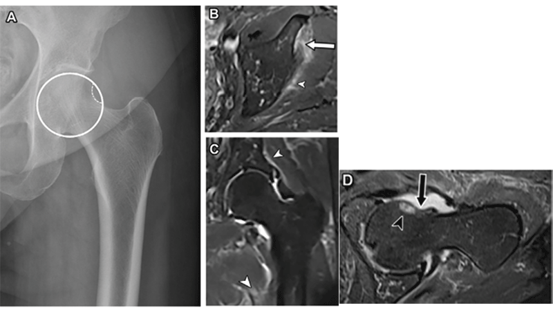
Iliofemoral ligament partial tear in a 41-year-old woman who presented with severe left hip pain following stair climbing 2 weeks earlier. Frontal pelvis radiograph (A) shows the cliff sign on the left hip, a steep drop-off, and loss of normal sphericity of the lateral femoral head (circle in A). Axial (B), coronal (C), and axial oblique (D) T2-weighted fat-suppressed MR images show signs of previous anterior hip subluxation, marked by partial tearing of the iliofemoral ligament at the acetabulum insertion site (arrow in B), as well as fluid leakage into the periarticular and muscle-adipose planes (arrowheads in B, C). Also note the divot sign (arrow in D), an indentation on the anterior surface of the femoral head, and contusional femoral head edema (arrowhead in D).
https://doi.org/10.1148/rg.240134 ©RSNA 2025
AI Segments Brain Bleeds on CT

Intracerebral hemorrhage (ICH) and related conditions like perihematomal edema (PHE) and intraventricular hemorrhage (IVH) require precise imaging to guide urgent treatment decisions and predict outcomes. Current manual methods are slow and often inaccurate, risking delays or unnecessary interventions.
Faster, more accurate diagnosis and treatment planning for brain hemorrhages are needed to improve outcomes and reduce delays in critical care.
In a study published in Radiology Advances, Jawed Nawabi, MD, MHBA, MSc, from Charité Universitätsmedizin Berlin, and colleagues aimed to evaluate an AI model (nnU-Net) for automated hemorrhage segmentation.
The researchers trained and validated a nnU-Net deep learning model using manually segmented CT scans from multiple stroke centers to assess its accuracy and speed in segmenting ICH, IVH and PHE.
“nnU-Net enables reliable, time-efficient segmentation of ICH, PHE and IVH, validated across multicenter, multivendor datasets of spontaneous ICH, showing potential to enhance clinical workflows,” the authors conclude.
Read the full article, “Cross-institutional automated multilabel segmentation for acute intracerebral hemorrhage, intraventricular hemorrhage, and perihematomal edema on CT.”
A Legacy of Science and Innovation
RSNA peer-reviewed journals have long been recognized as a trusted source for groundbreaking radiological research that moves medicine forward.
Even as the field progresses, the foundational knowledge contained within back issues of our journals offers critical context for interpreting current research and serves as an essential resource for learning.
To provide easy access to articles that include historical perspectives, cited references to data, methods, rare case studies and observations, RSNA created two special collections:
The Radiology Legacy Collection is a digital archive of nearly 100 years of articles, images and vintage advertisements from RSNA’s premier publication.
The RadioGraphics Legacy Collection brings together almost 40 years of high-quality educational articles, monograph issues and curated study guides.
Both collections are available free to RSNA members and non-member individual subscribers with an active subscription. Institutions may purchase the collections for a fee. Learn more now.
Peer Reviewers Needed
Help advance radiologic science and support your specialty— become a peer reviewer for RSNA journals. As a reviewer, you’ll play a vital role in maintaining the quality and integrity of published research while gaining insights into the latest advancements.
Your expertise can make a meaningful impact. Volunteer your time and talent to shape the future of radiology. Learn more now.

